
Fused image moldalities guidance

a moldalization and image technology, applied in the field of medical imaging, can solve the problems that the resolution of images may also affect the quality of registration, and the intensity of objects in images does not necessarily correspond, so as to achieve the effect of affecting the quality of registration

Inactive Publication Date: 2011-07-21
EIGEN INC
View PDF1 Cites 135 Cited by
  • Summary
  • Abstract
  • Description
  • Claims
  • Application Information

AI Technical Summary

Benefits of technology

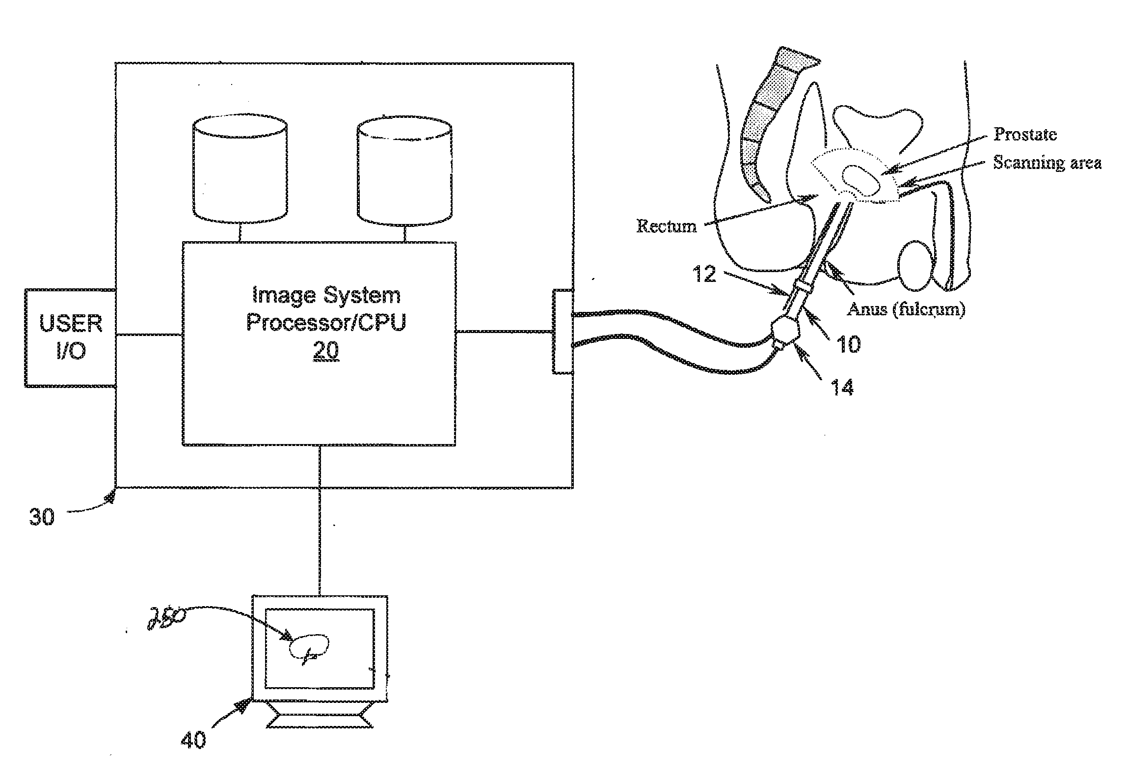
The term fusion is sometimes used to define the process of registering two images that are acquired via different imaging modalities. The present inventors have recognized that registration/fusion of images obtained from different modalities creates a number of complications. This is especially true in soft tissue applications where the shape of an object in two images may change between acquisitions of each image. Further, in the case of prostate imaging the frame of reference (FOR) of the acquired images is typically different. That is, multiple MRI volumes are obtained in high resolution transverse, coronal or sagittal planes respectively. These planes are usually in rough alignment with the patient's head-toe, anterior-posterior or left-right orientations. In contrast, TRUS images are often acquired while a patient lays on his side in a fetal position...

Problems solved by technology

A further difficulty with these different modalities is that the intensity of objects in the images do not necessarily correspond.
Finally, the resolution of the images may also impact registration quality.

Method used


Image

  • Fused image moldalities guidance
  • Fused image moldalities guidance
  • Fused image moldalities guidance

Examples


Embodiment Construction

Reference will now be made to the accompanying drawings, which assist in illustrating the various pertinent features of the present disclosure. The following description is presented for purposes of illustration and description.

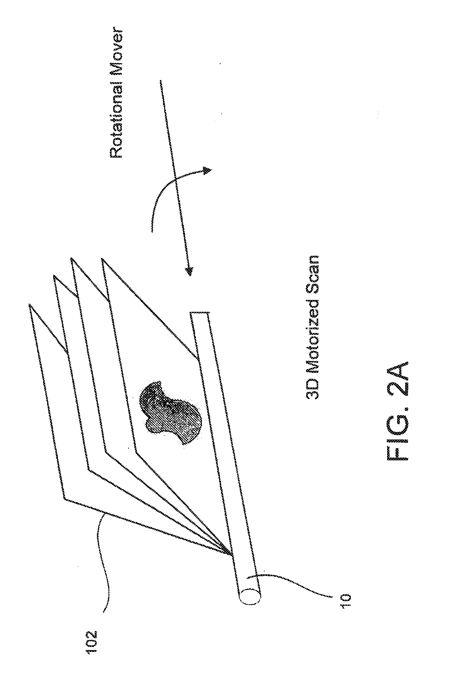
Disclosed herein are systems and methods that allow for registering images acquired from different imaging modalities (e.g., multimodal images) to a common frame of reference (FOR). In this regard, one or more images may be registered during, for example, an ultrasound guided procedure to provide enhanced patient information. Such registration of multimodal images is sometimes referred to as image fusion. In the application disclosed herein, a pre-acquired MRI image(s) of a prostate of a patient and a real-time TRUS image (e.g., 3D TRUS volume) of the prostate are registered such that information present in the MRI image(s) may be displayed in the FOR of the TRUS image to provide additional information that may be utilized for guiding a medical procedure on / a...


PUM


Abstract

An improved system and method (i.e. utility) for registration of medical images is provided. The utility registers a previously obtained volume (s) onto an ultrasound volume during an ultrasound procedure to produce a multimodal image. The multimodal image may be used to guide a medical procedure. In one arrangement, the multimodal image includes MRI information presented in the framework of a TRUS image during a TRUS procedure.

Description

FIELDThe present disclosure pertains to the field of medical imaging, and more particularly to the registration of multiple medical images to allow for improved guidance of medical procedures. In one application, multiple medical images are coregistered into a multimodal image to aid urologists and other medical personnel in finding optimal target sites for biopsy and / or therapy.BACKGROUNDMedical imaging, including X-ray, magnetic resonance (MR), computed tomography (CT), ultrasound, and various combinations of these techniques are utilized to provide images of internal patient structure for diagnostic purposes as well as for interventional procedures. One application of medical imaging (e.g., 3-D imaging) is in the detection and / or treatment of prostate cancer. According to the National Cancer Institute (NCI), a man's chance of developing prostate cancer increases drastically from 1 in 10,000 before age 39 to 1 in 45 between 40 to59 and 1 in 7 after age 60. The overall probability ...

Claims


Application Information

Patent Timeline
IPC IPC(8): A61B5/055A61B8/00
CPCA61B5/055A61B8/12A61B8/4461A61B8/483A61B8/5238G06T2207/30081G06T7/0083G06T7/0097G06T2207/10088G06T2207/10136G06T7/0028A61B8/4254G06T7/33G06T7/12G06T7/174
Inventor KUMAR, DINESHNARAYANAN, RAMKRISHNAN
Owner EIGEN INC
Learn More