
Image processing method for estimating a brain shift in a patient

a brain shift and image processing technology, applied in the field of computer-aided neurosurgery, can solve the problems of considerable impairment of neuronavigation, and achieve the effect of rapid, robust and precise respons

Inactive Publication Date: 2011-10-20
UNIV JOSEPH FOURIER +1
View PDF9 Cites 11 Cited by
  • Summary
  • Abstract
  • Description
  • Claims
  • Application Information

AI Technical Summary

Benefits of technology

[0013]The overall shift calculated in this way reveals the preoperative data and rectifies the initial surgical planning. The invention provides the surgeon with a rapid, robust and precise response and gives him the possibility of validating the pertinence of the calculated shift prior to any planning modification.

Problems solved by technology

Due to this brain shift, the preoperative data no longer correspond to reality, and neuronavigation is considerably compromised as a result.

Method used


Image

  • Image processing method for estimating a brain shift in a patient
  • Image processing method for estimating a brain shift in a patient
  • Image processing method for estimating a brain shift in a patient

Examples


Embodiment Construction

[0035]An embodiment of the process according to the invention will now be described in greater detail.

[0036]General Principle of the Invention

[0037]As explained previously, precise location of the surgical target is essential for reducing the morbidity during surgical exercises of a cerebral tumour. When the dimensions of the craniotomy are substantial a shift in the soft tissue of the brain can occur during the intervention. Due to this brain shift, the preoperative data no longer correspond to reality, and neuronavigation is considerably compromised as a result. The present invention takes this shift into consideration and calculates a rectified position of the anatomical structures of the brain to locate the auxiliaries.

[0038]Prior to a surgical operation on the brain of a patient, a magnetic resonance angiography (MRA) of the brain of the patient can be acquired. During the intervention, as a result of a significant brain shift the surgeon carries out echographic Doppler scannin...


PUM


Abstract

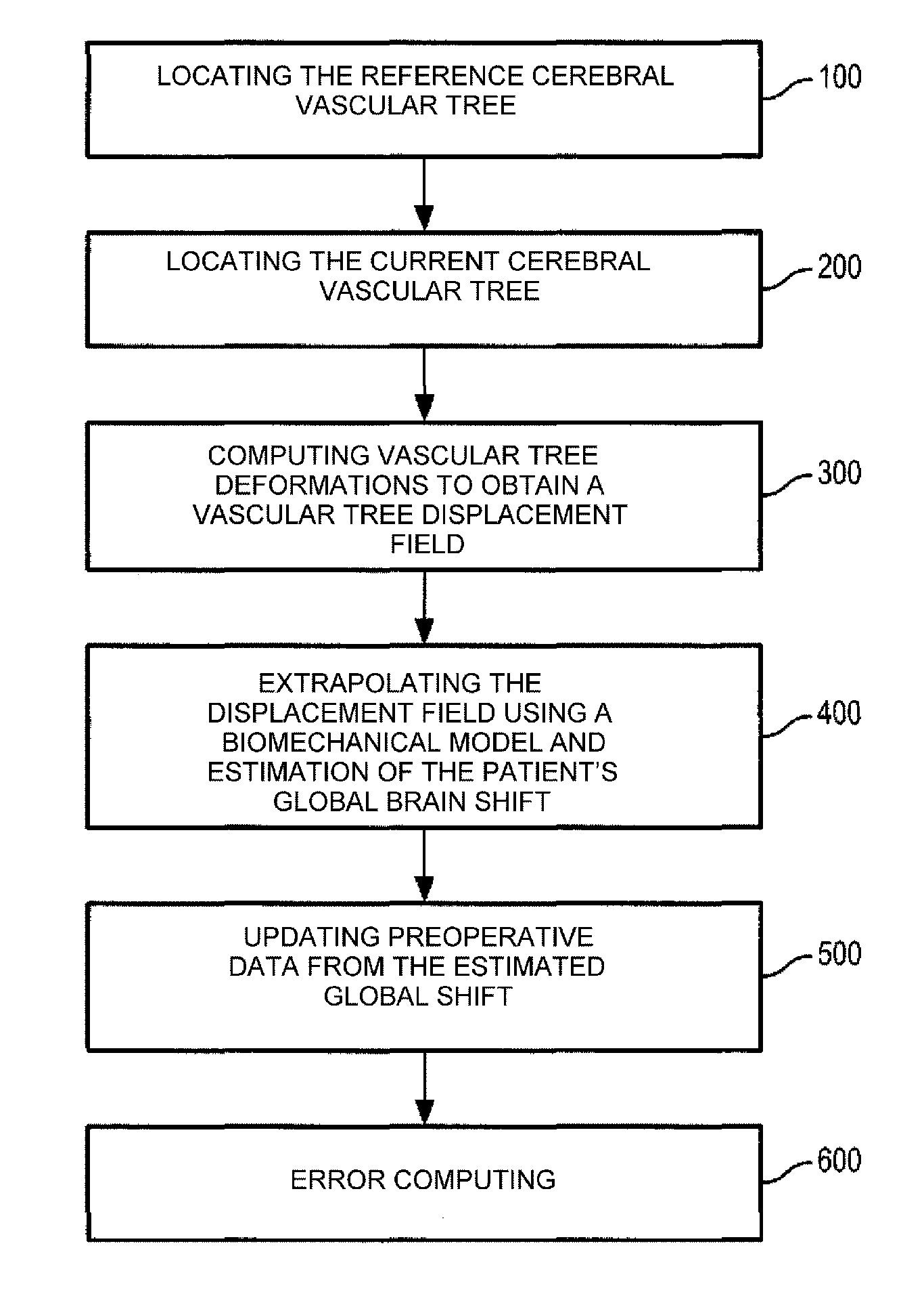
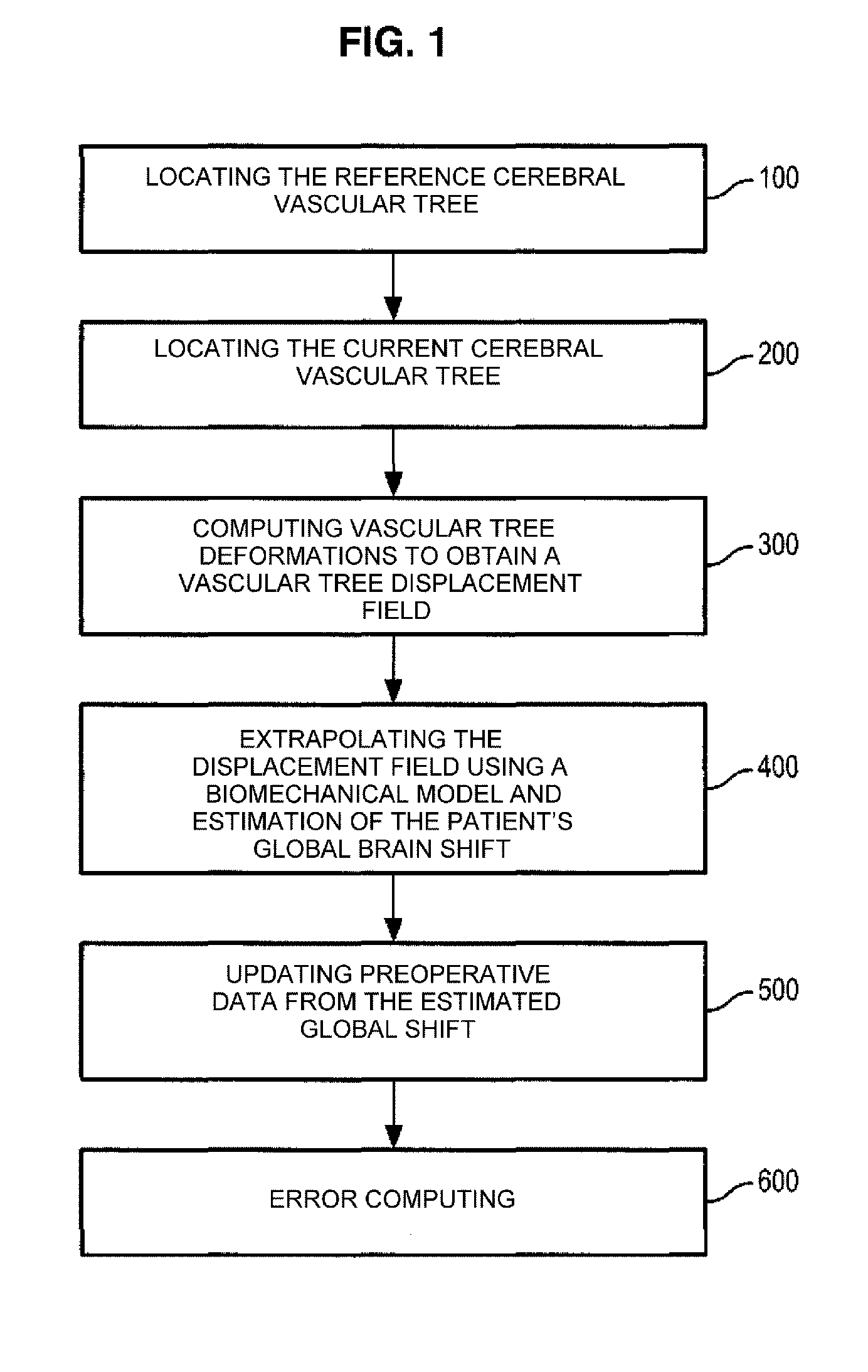
The disclosure relates to an image processing method for estimating a brain shift in a patient, the method involving: the processing of a three-dimensional image of the brain of a patient, acquired before a surgical operation, in order to obtain a reference cerebral arterial tree structure of the patient; the processing of three-dimensional images of the brain of the patient, acquired during the operation, in order to at least partially reconstitute a current cerebral arterial tree structure of the patient; the determination from the combination of the reference and current cerebral arterial tree structures, of a field of shift of the vascular tree representing the shift of the current vascular tree in relation to the reference vascular tree; the application of the determined field of shift of the vascular tree to a biomechanical model of the brain of the patient in order to estimate the brain shift of the patient; and the generation, from the estimated brain shift, of at least one image of the brain of the patient, in which the brain shift is compensated.

Description

CROSS-REFERENCE TO RELATED APPLICATIONS[0001]This application is a National Phase Entry of International Application No. PCT / EP2009 / 062826, filed on Oct. 2, 2009, which claims priority to French Patent Application Serial No. 0856712, filed on Oct. 3, 2008, both of which are incorporated by reference herein.FIELD OF THE INVENTION[0002]The present invention relates to the field of computer-assisted neurosurgery, and more particularly a system and method for precise location of a target.BACKGROUND[0003]Precise location of a target is essential for reducing the morbidity rate during a surgical operation involving cerebral tumour ablation. During a surgical operation on the brain, a shift in cerebral tissues (or “brain shift” according to English terminology) can occur after an opening has been made in the cranium of the patient. This shift can be associated with physical phenomena (gravity, loss of cephalorachidian liquid, action by the neurosurgeon, etc.) or with physiological phenomen...

Claims


Application Information

Patent Timeline
IPC IPC(8): A61B5/055A61B6/00A61B8/08
CPCG06T7/0038A61B8/0808G06T2207/30016G06T2207/10072G06T7/38
Inventor BUCKI, MAREKPAYAN, YOHAN
Owner UNIV JOSEPH FOURIER
Learn More