Increasing the plasticity of stem cells
a stem cell and plasticity technology, applied in the field of stem cell plasticity, can solve the problems of increasing the culture time required between the removal of the cells, increasing the chance of tumourigenesis, and requiring complicated cell genetic manipulation, so as to enhance the pluripotency of cells
- Summary
- Abstract
- Description
- Claims
- Application Information
AI Technical Summary
Benefits of technology
Problems solved by technology
Method used
Image
Examples
example 1
Spheroid Production Method Development and Characterisation
Materials and Methods
[0083]A mouse osteogenic cell line (C3H10t1 / 2) was used for method development. Cells were trypsinised and seeded at a specific cellular density into non adherent 96 well U shaped plates. Cells were resuspended at a density of 3×10⁴, 6×10⁴, 1.2×10⁵, and 2.4×10⁵ in 200 μl of Dulbecco's modified Eagles medium containing 100 U / ml penicillin and 100 μg / ml streptomycin, 15% FBS, and 0.25% methyl cellulose. Spheroids were incubated at 37° C. in 5% CO2 in 95% air with 90% humidity. Spheroids were cultured for 1-7 days and images captured using a light microscope.
Results
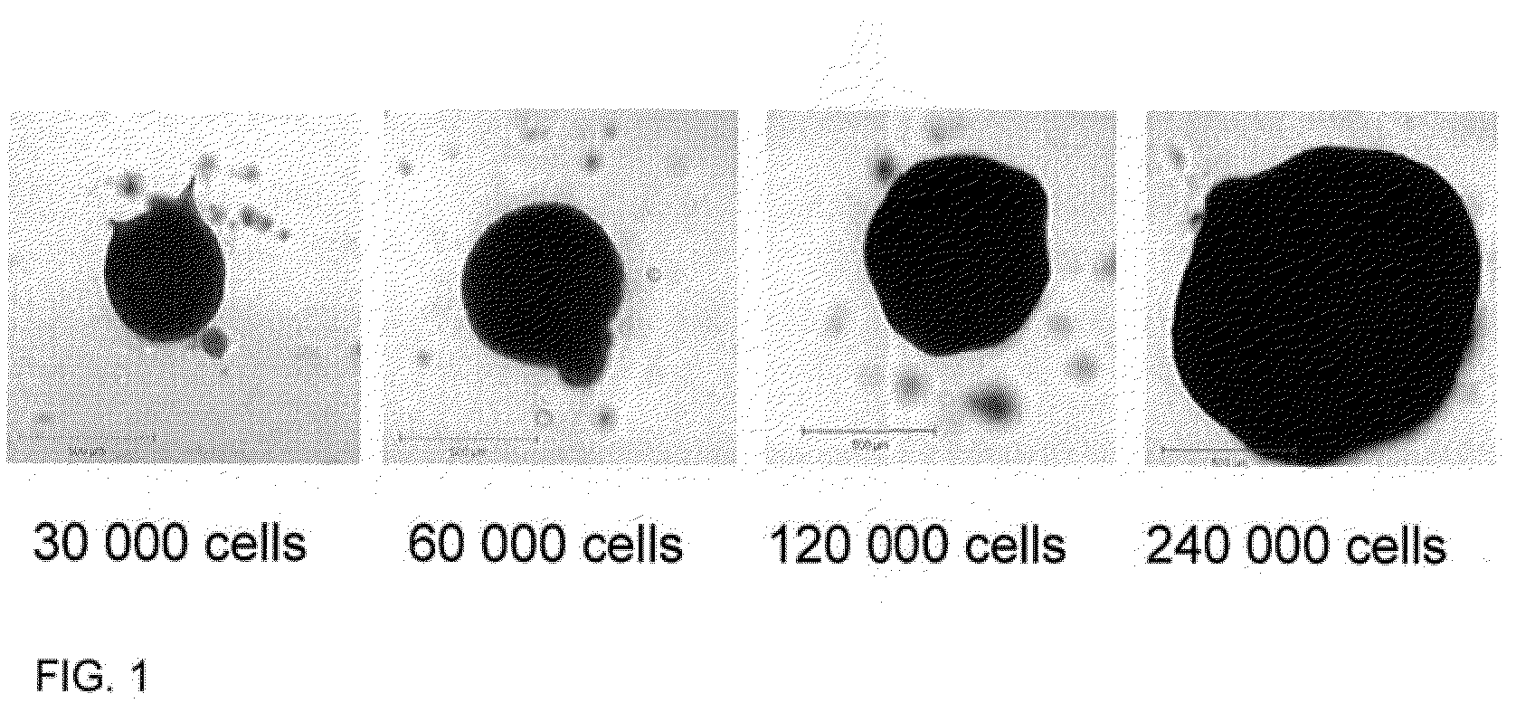
[0084]FIG. 1 represents images of spheroids produced using this method, with increasing numbers of cells / spheroid shown.
[0085]The method outlined above can be used to produce regular shaped and sized spheroids and as such is a reproducible and reliable model.
example 2
Immature Non-Embryonic Cells Derived from Human MSCs
Materials and Methods
i) Cell Culture Medium
[0086]Dulbecco's modified Eagles medium containing 100 U / ml penicillin and 100 μg / ml streptomycin, 15% FBS, and 0.25% methyl cellulose. Cells were incubated at 37° C. in 5% CO2 in 95% air with 90% humidity.
ii) Isolation of Mesenchymal Stem Cells from Femoral Heads
[0087]Femoral heads from routine hip replacements were obtained. The trabecular bone was removed from the centre of the femoral head and transferred Dulbecco's modified Eagles medium (DMEM) containing 100 U / ml penicillin and 100 μg / ml streptomycin. The trabecular bone was minced with scissors, fragments allowed to settle and the media transferred to another tube. This was repeated another two times and the bone fragments vortexed before transferring the media.
[0088]This cell suspension was centrifuged at 500 g for 5 minutes and the pellet resuspended in 16 ml of DMEM. This suspension was then passed through a 70 μm cell sieve, to ...
example 3
Determination of the Expression of Embryonic Transcripts Following Culture of Human Dermal Fibroblasts in a 3D Environment
[0103]In order to determine whether or not the increase in embryonic transcripts were as a result of the 3D environment, the cell type or a combination of both, human dermal fibroblasts were cultured in 3D and the expression of Oct4, Nanog, SOX2 and Rex1 was determined.
Materials and Methods
[0104]Method as in Example 1 and 2.
Results
[0105]As FIG. 6 illustrates the expression of both Oct4 and Nanog were up-regulated in the 3D cultures, however the expression of Rex1 and SOX2 could not be detected. Such results suggest that the 3D culture does play an important role in the up-regulation of embryonic transcripts but that the effect is greater when more primitive cells (MSCs) are used.
PUM
| Property | Measurement | Unit |
|---|---|---|
| density | aaaaa | aaaaa |
| humidity | aaaaa | aaaaa |
| humidity | aaaaa | aaaaa |
Abstract
Description
Claims
Application Information
 - R&D
- Intellectual Property
- Life Sciences
- Materials
- Tech Scout
- Unparalleled Data Quality
- Higher Quality Content
- 60% Fewer Hallucinations
Browse by: Latest US Patents, China's latest patents, Technical Efficacy Thesaurus, Application Domain, Technology Topic, Popular Technical Reports.
© 2025 PatSnap. All rights reserved.Legal|Privacy policy|Modern Slavery Act Transparency Statement|Sitemap|About US| Contact US: help@patsnap.com


