
Liver lesion segmentation

a liver tumor and segmentation technology, applied in the field of diagnostic imaging, can solve the problems of not being able to accurately track the volume change of irregularly shaped tumors, being unsuitable for the overall workflow requirements, and being tedious and time-consuming, so as to improve the segmentation performance, improve the segmentation effect, and reduce the likelihood of segmentation leakage

Active Publication Date: 2011-12-29
PHILIPS HEALTHCARE INFORMATICS INC
View PDF4 Cites 14 Cited by
  • Summary
  • Abstract
  • Description
  • Claims
  • Application Information

AI Technical Summary

Benefits of technology

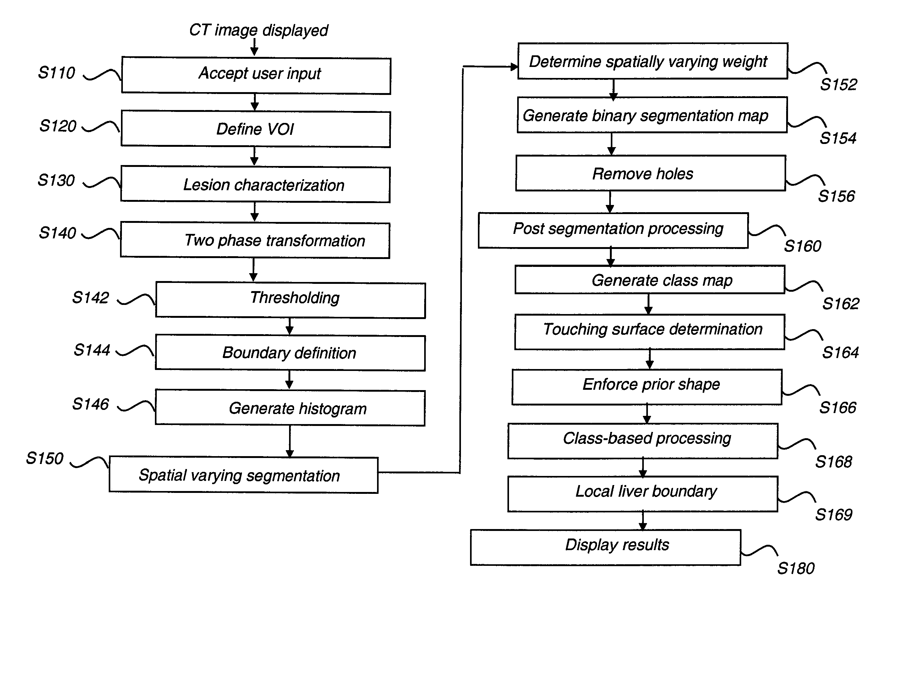
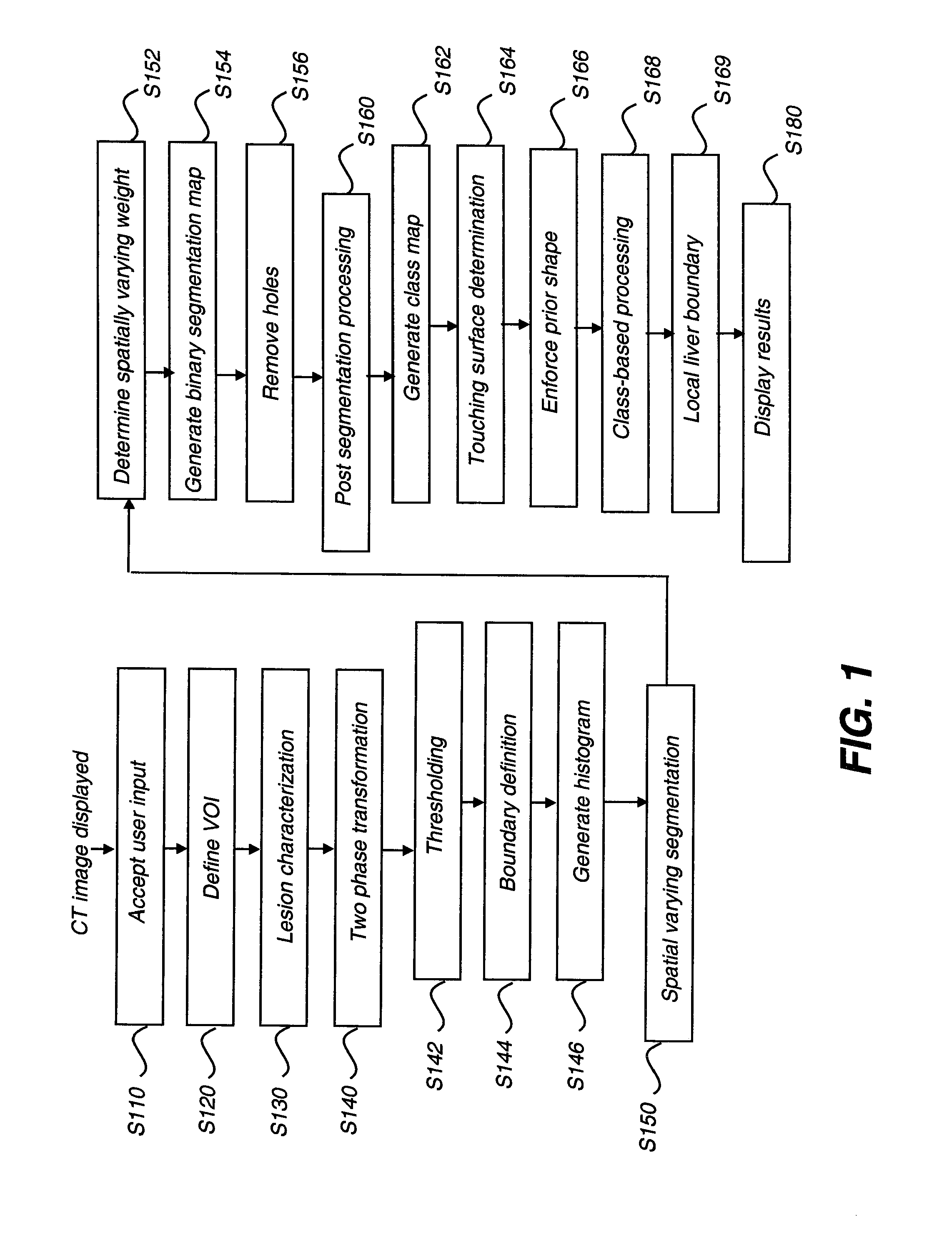
[0009]An object of the present invention is to provide a method for improved segmentation of lesions of the liver and similar organs. The method of the present invention is semi-automatic, segmenting a lesion according to initial input from the radiologist. Measures are taken to improve segmentation performance and to reduce the likelihood of segmentation leakage.

Problems solved by technology

These linear measurements, obtained from manual radiologist mark-up, are of some value but are generally not sufficient for accurately tracking the volume change of irregularly shaped tumors.
This process is a tedious, time-consuming task and is often poorly suited to the overall workflow requirements of the radiologist.
In addition, the results of manual segmentation can be highly dependent upon the observer and are often not reproducible.
Unenhanced CT images are generally insufficient for detection of lesions because there is very little difference in attenuation values between the lesion and parenchyma, or surrounding tissue, in the liver.
Once detected, segmentation of liver lesions is a challenging task because of the large variability of intensity values, in Hounsfield units (HU), between the lesion and parenchyma in the liver.
One persistent problem, due to the properties of the parenchyma and surrounding tissues within and outside of the liver, relates to leakage, a segmentation phenomenon in which the boundaries of an identified lesion may not be clearly distinguishable from nearby tissues, causing the segmentation algorithm to mistakenly categorize non-lesion tissue as belonging to the lesion.

Method used


Image

  • Liver lesion segmentation
  • Liver lesion segmentation
  • Liver lesion segmentation

Examples


Embodiment Construction

[0022]The following is a detailed description of the preferred embodiments of the invention, reference being made to the drawings in which the same reference numerals identify the same elements of structure in each of the several figures.

[0023]In typical applications, a computer or other type of general-purpose or dedicated logic processor is used for obtaining, processing, and storing image data as part of the CT system, along with one or more displays for viewing image results. A computer-accessible memory is also provided, which may be a non-volatile memory storage device used for longer term storage, such as a device using magnetic, optical, or other data storage media. In addition, the computer-accessible memory can comprise an electronic memory such as a random access memory (RAM) that is volatile, used for shorter term storage, such as employed to store a computer program having instructions for controlling one or more computers to practice the method according to the present...


PUM


Abstract

A method for segmenting a first object from a bounding object within a digital image identifies pixels between first and second endpoint pixels. A volume of interest is defined spanning at least a portion of the object. Pixels within the volume of interest are partitioned into a first subset of pixels that are neither the first object nor bounding object pixels and second subset of pixels that are not contained in the first subset. A spatially varying two-phase segmentation process segments the first object from the bounding object within the second subset of pixels according to the location of each subject pixel within the second subset relative to pixels in the first subset of pixels. The first object segmentation is refined according to the subject pixel location relative to the segmented bounding object and to the first subset of pixels. A segmented image is formed according to the refined segmentation.

Description

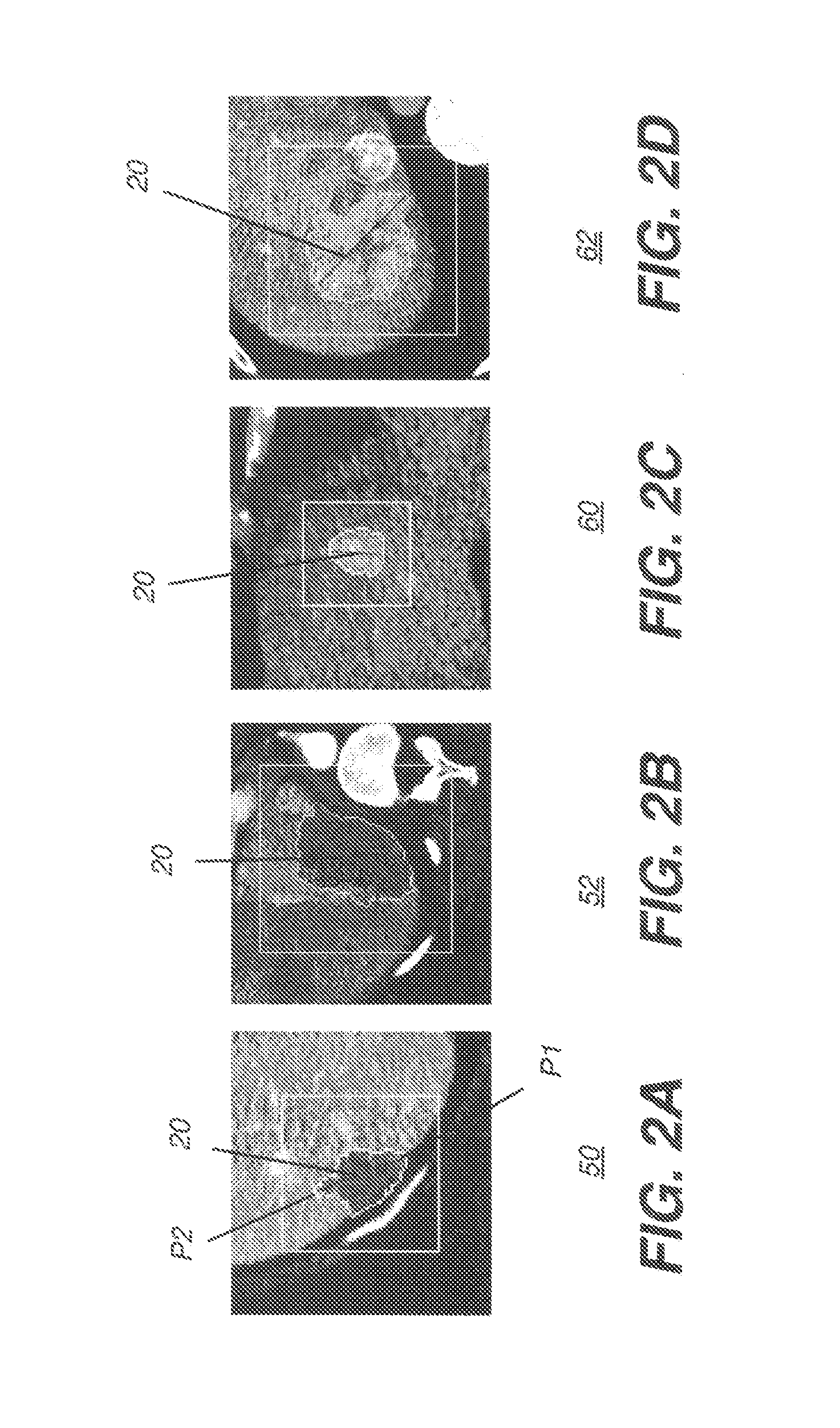
FIELD OF THE INVENTION[0001]The invention relates generally to the field of diagnostic imaging and more particularly relates to a method for assisted segmentation of liver tumors, particularly in computed tomography images.BACKGROUND OF THE INVENTION[0002]Identification of potentially cancerous lesions in computed tomography (CT) and magnetic resonance imaging (MRI) studies is a common task of radiologists. An aspect of this task relates to measurement of lesion volume and density, both important in assessing the temporal response of a tumor to treatment.[0003]RECIST (Response Evaluation Criteria In Solid Tumors) is a set of published rules that define when cancer patients improve (“respond”), stay the same (“stable”) or worsen (“progression”) during treatments. Typically, a uni-dimensional measurement, RECIST criteria, which relates to the longest diameter of the tumor, is used as a type of surrogate for 3D volume measurement. These linear measurements, obtained from manual radiolo...

Claims


Application Information

Patent Timeline
IPC IPC(8): G06K9/34G06K9/00
CPCG06K9/6207G06T7/0081G06T7/0089G06T2207/30056G06T2207/20116G06T2207/20148G06T2207/20168G06T2207/20104G06T7/11G06T7/149G06T7/136G06V10/755
Inventor SIMON, RICHARD A.
Owner PHILIPS HEALTHCARE INFORMATICS INC
Learn More