Endoscopic image processing device, method and program

a processing device and endoscopic technology, applied in image analysis, image enhancement, medical science, etc., can solve the problems of inability to apply various known analysis methods and image display methods, such as volume rendering and display methods, to achieve more accurate imaging diagnosis

Inactive Publication Date: 2012-03-01
FUJIFILM CORP
View PDF4 Cites 30 Cited by
  • Summary
  • Abstract
  • Description
  • Claims
  • Application Information

AI Technical Summary

Benefits of technology

[0034]It should be noted that endoscopic images have a nature that an area near to the imaging unit, such as a CCD camera, of the endoscope appears larger, and an area far from the imaging unit appears smaller. Therefore, the number of pixels representing the area far from the imaging unit is smaller than the number of pixels representing the area near to the imaging unit. In the generated volume data, therefore, a position corresponding to the area far from the imaging unit in the endoscopic image has a smaller number of positions on the endoscopic image associated therewith than those associated with a position corresponding to the area near to the imaging unit in the endoscopic image. In that case, there may be only sparse positions in the volume data that correspond to the endoscopic image. Therefore, by calculating and associating a plurality of three-dimensional positions in a certain spatial range for and with each pixel in an area in the endoscopic volume data that is far from the imaging unit and has smaller number of positions on the endoscopic image associated therewith, appropriate interpolation of pixel values in the area of the endoscopic volume data that has sparse corresponding pixels on the endoscopic image can be achieved. This minimizes such a situation that there are sparse positions in the volume data associated with the endoscopic image, and the volume data with each pixel reflecting an appropriate pixel value can be generated.
[0035]In the case where the endoscopic image processing device according to the invention includes the reconstructed endoscopic image generating means, and the reconstructed endoscopic image generated by the reconstructed endoscopic image generating means is a stretched development image or a stretched cross-sectional image of the anatomical structure, image observation that cannot be achieved with conventional endoscopic images can be achieved, thereby providing a support for more accurate imaging diagnosis.

Problems solved by technology

However, while an endoscopic image provides an image that clearly represents colors and textures of the interior of an anatomical structure captured with an image pickup device, such as a CCD, the interior of the anatomical structure is shown in a two-dimensional image, and therefore it is difficult to understand which position in the anatomical structure the image shows, and it is impossible to obtain information of the depth direction to reach a desired part.
However, while the methods disclosed in Patent Documents 1 to 3 provide understanding of the position of the endoscope in an anatomical structure, it is impossible to apply various known analysis methods and image display methods, such as volume rendering and a display method for displaying a stretched development view of a tubular structure, unlike volume data acquired through tomographic imaging with a CT apparatus, or the like.
Further, while the method disclosed in Patent Document 4 provides three-dimensional understanding of the interior of an anatomical structure, it does not convert the endoscopic images into volume data.
Therefore, unlike volume data, it is impossible to use various image analysis methods and image display methods with the endoscopic images.

Method used


Image

  • Endoscopic image processing device, method and program
  • Endoscopic image processing device, method and program
  • Endoscopic image processing device, method and program

Examples


first embodiment

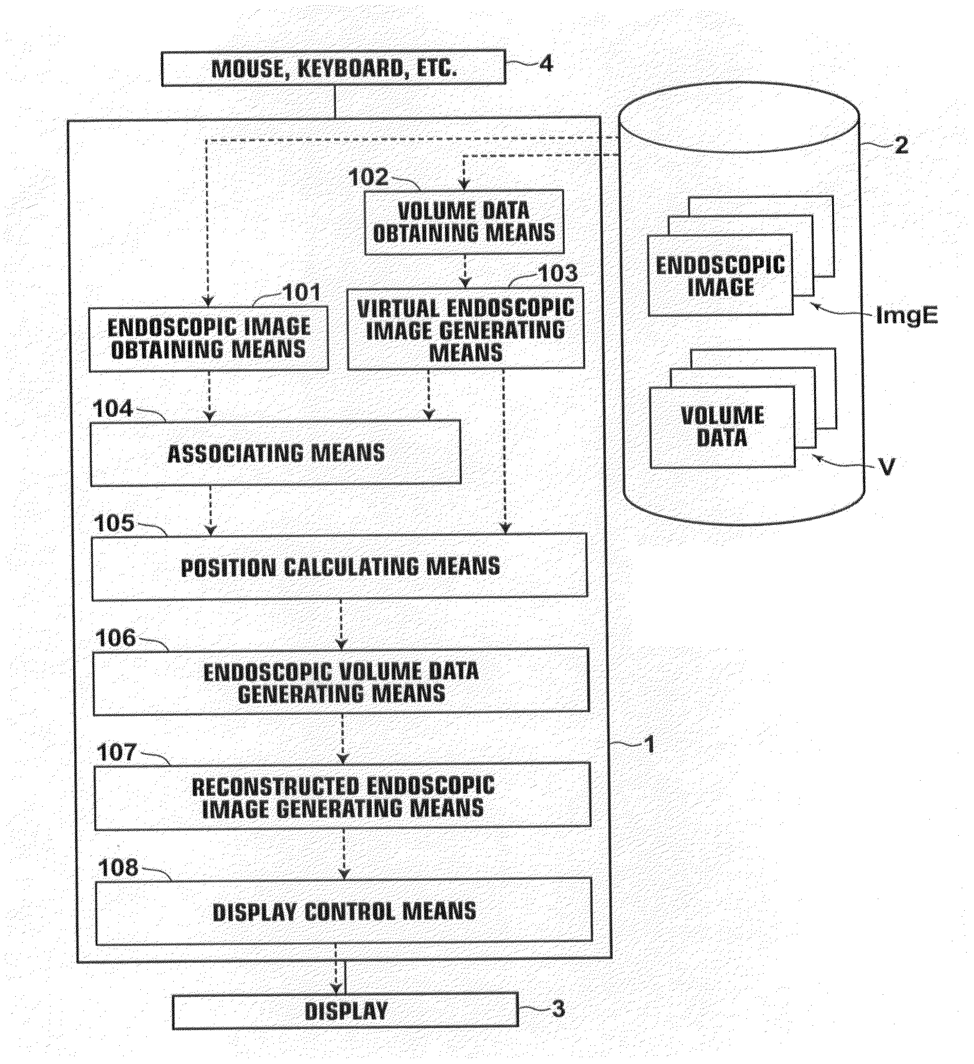
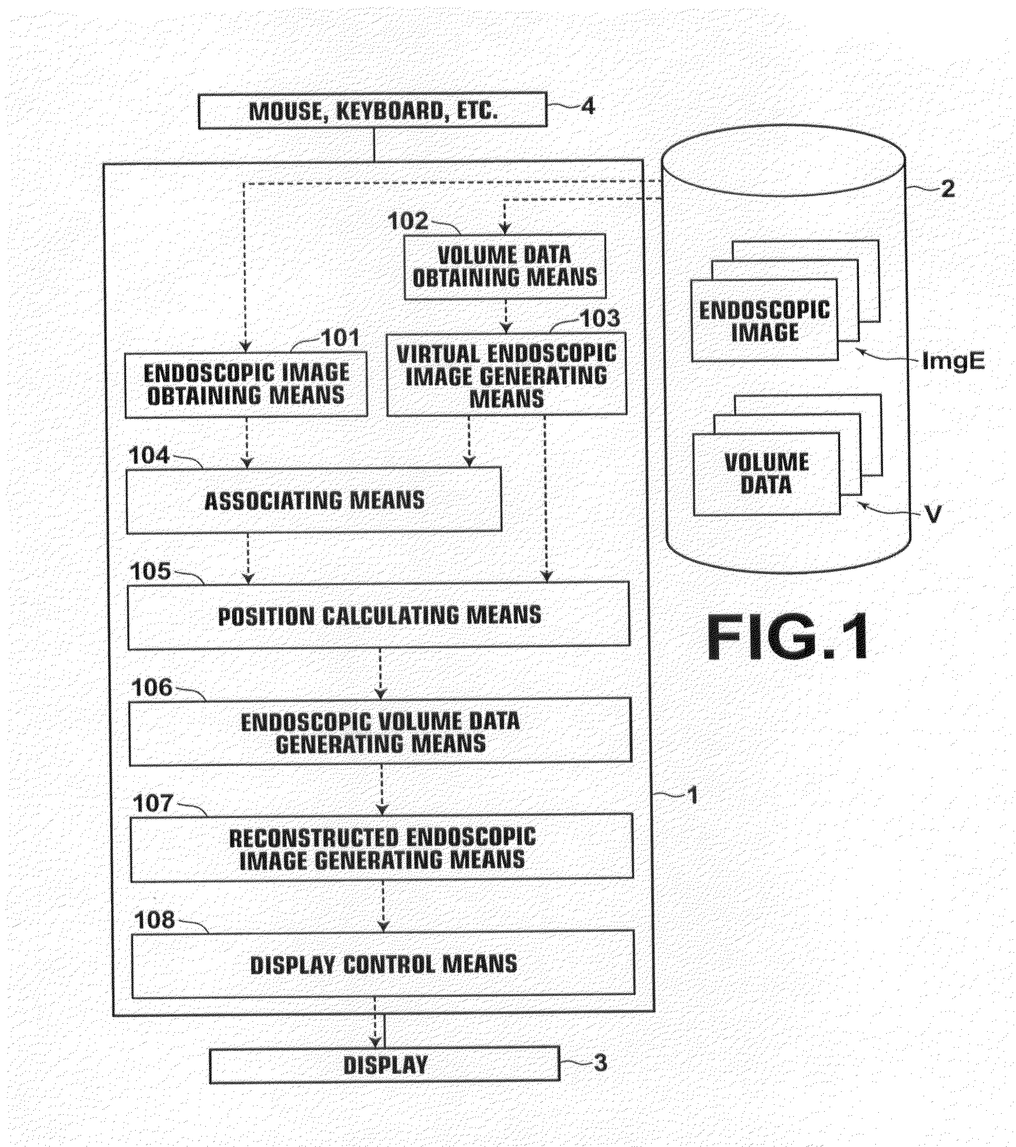
[0105]An area in the endoscopic volume data VE formed by three-dimensional positions that are associated with none of the pixels of the endoscopic image is referred to as the “unassociated area”. For example, the endoscopic volume data VE, where the pixel values of certain pixels are reflected in the three-dimensional positions, is generated by calculating the three-dimensional positions corresponding to the pixels forming the endoscopic images. Therefore, it is impossible to associate an unimaged area in the anatomical structure, such as an area which is not captured in the endoscopic images due to a bumpy shape, such as a polyp, or a curved shape in the anatomical structure, with the three-dimensional positions of the endoscopic volume data VE. Further, there are cases where some pixels on the endoscopic images are not associated with the three-dimensional positions, such as the case where the endoscopic image processing of the invention is applied to only a part of each endoscop...

second embodiment

[0106]In this second embodiment, the unassociated area extracting means 110 detects, from pixels forming the anatomical structure, pixels having pixel values that are not changed from the initial values, and extracts an area formed by the extracted pixels as the unassociated area Ru.

[0107]Further, the medical image generating means 109 generates a medical image ImgRv from the volume data V obtained from the storage 2. In the second embodiment, a volume rendered image is generated by volume rendering as the medical image ImgRv. The medical image ImgRv may be any image that can be generated from the volume data V, and typical examples thereof include images generated by volume rendering and surface rendering.

[0108]FIG. 6 is a flow chart illustrating the flow of processing according to the second embodiment. FIG. 7 shows an example of markings M indicating the unassociated areas Ru displayed in an identifiable manner on the medical image ImgRv by the processing of the second embodiment...


PUM


Abstract

Endoscopic images and virtual endoscopic images are obtained. Then, an endoscopic image captured at a predetermined position of the anatomical structure is extracted from the obtained endoscopic images, and a comparative virtual endoscopic image virtually generated as if it is captured at a position corresponding to the predetermined position is extracted from the obtained virtual endoscopic images, and the extracted images are associated with each other. A three-dimensional position corresponding to each pixel forming the endoscopic image captured at the predetermined position is calculated based on a three-dimensional position of each pixel forming the comparative virtual endoscopic image. Then, volume data is generated from the endoscopic image captured at the predetermined position based on a pixel value of each pixel forming the endoscopic image and the three-dimensional position calculated for each pixel.

Description

BACKGROUND OF THE INVENTION[0001]1. Field of the Invention[0002]The present invention relates to a technique to process an endoscopic image during surgery or examination with an endoscope being inserted in an anatomical structure of a subject, and in particular to a technique to process an endoscopic image with using a virtual endoscopic image that represents an anatomical structure of a subject.[0003]2. Description of the Related Art[0004]In recent years, techniques to observe or treat an anatomical structure, such as the intestine or the bronchial tube, of a patient with using an endoscope are attracting attention.[0005]However, while an endoscopic image provides an image that clearly represents colors and textures of the interior of an anatomical structure captured with an image pickup device, such as a CCD, the interior of the anatomical structure is shown in a two-dimensional image, and therefore it is difficult to understand which position in the anatomical structure the image...

Claims


Application Information

Patent Timeline
Patent Type & Authority Applications(United States)
IPC IPC(8): A61B1/04
CPCG06T2207/10068G06T2207/10072G06T2207/20221G06T2219/2012G06T19/20G06T2207/10124G06T2210/41G06T7/0044G06T7/74
Inventor MIYAMOTO, MASAKI
Owner FUJIFILM CORP
Why Eureka
Try Eureka