Ultrasound Registration

a technology of ultrasound and registration, applied in the field of ultrasound registration, can solve the problems of inability to directly manipulate the ultrasound transducer, surgeons are often removed from patients, and standard visible-light cameras are limited by their inability to visualize subsurface anatomic features

Inactive Publication Date: 2012-03-22
THE UNIV OF BRITISH COLUMBIA
View PDF6 Cites 25 Cited by
  • Summary
  • Abstract
  • Description
  • Claims
  • Application Information

AI Technical Summary

Problems solved by technology

Standard visible-light cameras are limited by their inability to visualize subsurface anatomic features including vessels, suspicious lesions and organ or tumour boundaries.
In laparoscopic or robotic-assisted laparoscopic procedures, the surgeon is often removed from the patient and unable to directly manipulate the ultrasound transducer as needed for imaging during the procedure.

Method used


Image

  • Ultrasound Registration
  • Ultrasound Registration
  • Ultrasound Registration

Examples


Embodiment Construction

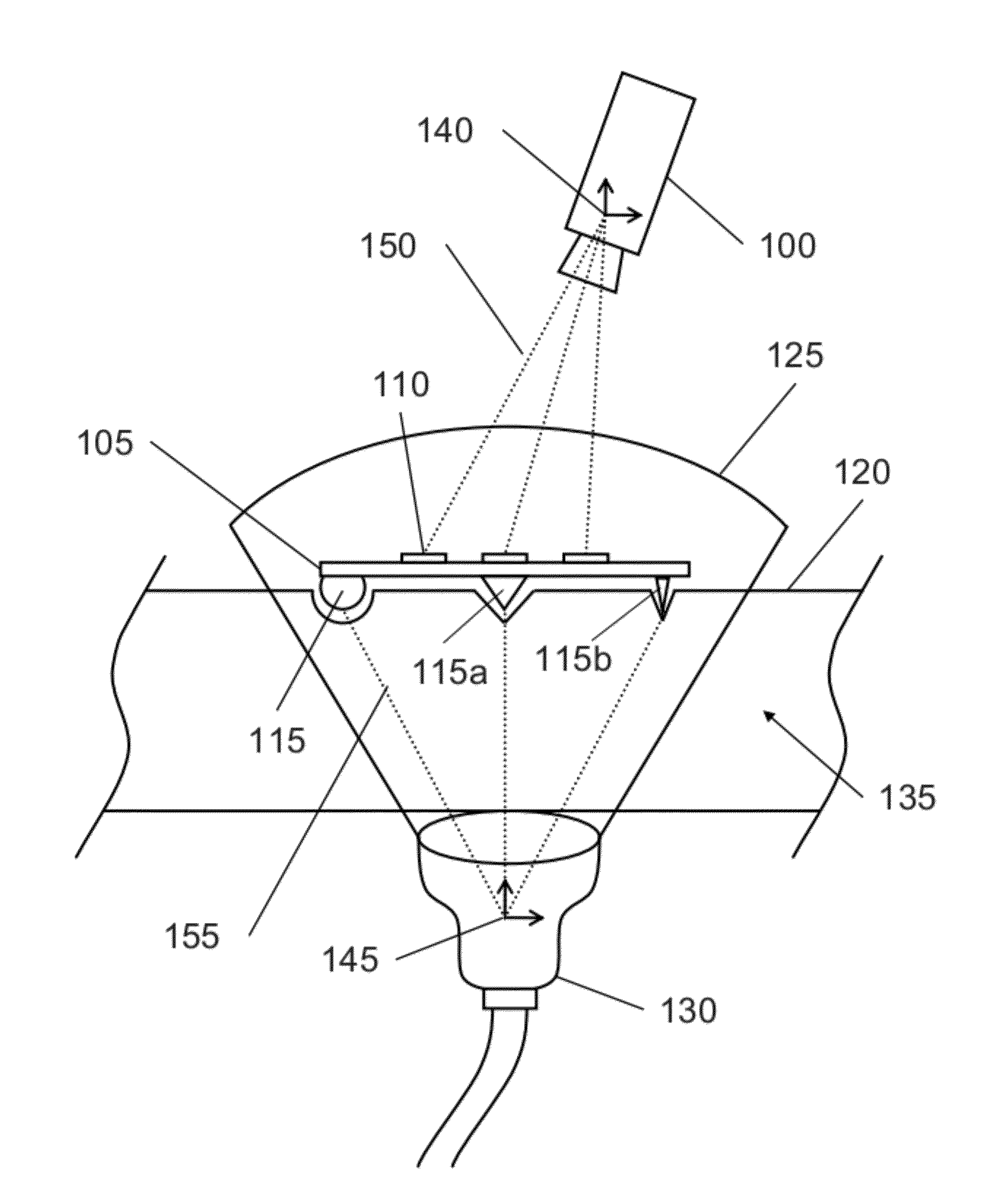
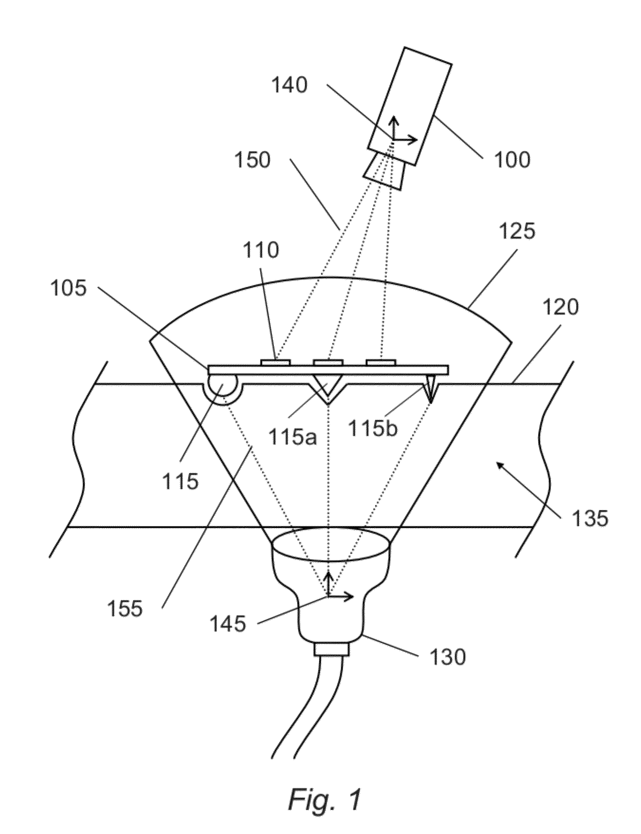
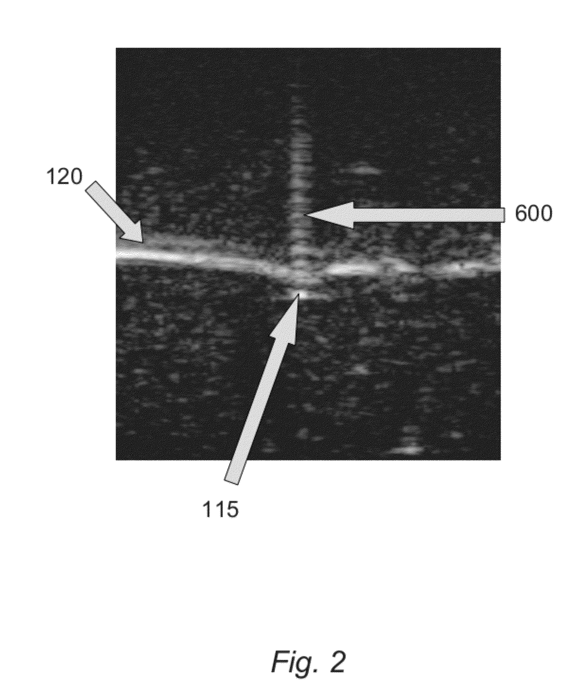
[0021]Throughout the following description, an ultrasound image feature refers to an aspect of an ultrasound image that can be detected and localized in an ultrasound image. The term surface is used for sake of illustration of the invention to describe the interface between tissue and air, but it may also describe the interface between tissue and fluid, and tissue and artificial tissue, including but not limited to soft rubbers. Ultrasound image features on a tissue surface may be created in ultrasound images by impressing, for example, a spherical ball bearing (referred to in this description as a surface fiducial), a needle tip, or the tip of a pointed object. Any other object that creates a distinct hyper-echoic or hypo-echoic image feature may also be used. Metal or any other material that generates strong reflections in ultrasound is preferred. Surfaces may be roughened or otherwise processed to contain ridges that strengthen the ultrasound echo signals they generate, as done, ...


PUM


Abstract

The present invention relates to a method and apparatus for registering three-dimensional ultrasound images to an external coordinate system, such as the coordinate system attached to a camera-based imaging system or the coordinate system used to define the movements of a surgical robot. The apparatus generally comprises a tool with markers that can be accurately and repeatably located in both the ultrasound and external coordinate systems. In the case of the surgical robot frame, this tool may be a component of the surgical robot itself, such as a portion of a surgical manipulator. The method generally comprises positioning the toot upon the surface of a portion of patient tissue, imaging the tool using the ultrasound, simultaneously imaging the tool using the camera system or monitoring the current mechanical configuration of the surgical robot, localizing corresponding tool elements in both frames, and finally using the corresponding elements as registration fiducials to solve the geometric transformation that relates the two frames.

Description

[0001]This application claims the benefit of U.S. Provisional Patent Application No. 61 / 344706, entitled ULTRASOUND TO CAMERA REGISTRATION, filed on Sep. 17, 2010, which is incorporated herein by reference.FIELD OF THE INVENTION[0002]The present invention relates to image guidance for medical interventions in general and three-dimensional (3D) ultrasound guidance for laparoscopic and robotic-assisted surgical procedures specifically.BACKGROUND OF THE INVENTION[0003]3D ultrasound has been used to create geometric models of tissue in a number of applications, including fetal and prostate imaging. In 3D ultrasound (for example, U.S. Pat. No. 5,454,371 “Method and System for Constructing and Displaying Three-Dimensional Images”) the ultrasound probe is swept over the target volume and the reflected ultrasound echoes are conveyed to a computer wherein successive two-dimensional images of the target volume are reconstructed to form a three-dimensional image of the target volume. Such a vo...

Claims


Application Information

Patent Timeline
Patent Type & Authority Applications(United States)
IPC IPC(8): A61B8/14
CPCA61B8/0841A61B19/2203A61B19/5244A61B2019/5289A61B2019/5255A61B2019/5276A61B2019/5287A61B2019/5251A61B34/20A61B34/30A61B2034/2051A61B2034/2055A61B2090/363A61B2090/364A61B2090/378
Inventor SALCUDEAN, SEPTIMIUYIP, MICHAEL CHAK LUENADEBAR, TROY KIEFERTROHLING, ROBERT NICHOLAS
Owner THE UNIV OF BRITISH COLUMBIA
Why Eureka
Try Eureka