Direct visualization robotic intra-operative radiation therapy applicator device

a radiation therapy and robotic technology, applied in the field of radiation cancer treatment, can solve the problems of difficult to obtain what is referred to as a clear margin, difficult to achieve the effect of clear margin, and difficult to achieve clear margin, etc., to achieve the effect of improving outcomes, and avoiding radiation therapy complications

Active Publication Date: 2012-01-10
SRIORT
View PDF29 Cites 132 Cited by
  • Summary
  • Abstract
  • Description
  • Claims
  • Application Information

AI Technical Summary

Benefits of technology

[0005]As this invention is intended to be used intra-operatively, surgeons skilled in the art of cancer surgery, together with radiation oncologists and medical physicists skilled in the art of using and delivering radiation treatments will use the invention cooperatively at the time of surgical removal of the tumor and at subsequent intervals as may be necessary to deliver radiation treatments intra-operatively as part of a planned surgical procedure to deliver curative doses of radiation to tumors. The invention, using imaging techniques such as ultrasound, MRI, CT, PET or PET / CT or some combination of medical imaging guidance, a priori or contemporaneously with the surgical procedure to guide and direct the radiation oncologist in the correct and accurate placement of the radiation field inside the patient and timing of tissue exposures to produce a curative dose of radiation without delivering doses to uninvolved tissues to minimize, to the greatest extent possible the complications associated with radiation treatment and delivery. The invention described herein will allow the operator to identify neoplastic tissue (benign or cancerous) of interest to the operator via medical imaging as described above, real time guidance via spatial depiction of the key anatomical landmarks at the time of insertion of the capsule for irradiation intra-operatively, realtime depiction in 3-dimensions on the imaging display system of the precise position of the applicator through the surgical robots' positioning reporting technologies and under direct visualization using visible light techniques and permit the operator to precisely position the intraoperative radiotherapy capsule in such a way, within the human body, using the surgical robotic manipulator arms under remote control of the robot by the physician, to deliver the proper type and exposure of radiation to the neoplastic tumors, thus enhancing the probability of curing and / or better managing the disease.

Problems solved by technology

Traditionally, intraoperative radiation therapy has been delivered via large, cumbersome linear accelerators and via injections of radioactive substances, both of which can cause substantial collateral damage and resultant morbidity and have not been shown to substantially improve outcomes.
A significant and longstanding problem with many cancers, such as ovarian cancer, is that upon resection (surgery), it is difficult to obtain what is referred to as a clear margin, or optimal debulking, that is a complete surgical removal of all cancer, including microscopic cancer.
As a result, residual cancer cells frequently remain, and may (and often do) break off from the primary cancer and migrate to other locations which are difficult to reach and destroy.
The metastatic cancer cells will then begin to grow using the local blood supply of the new site of involvement, eventually compromising organ function, and ultimately destroying the organ, frequently resulting in death.
Traditional external beam radiation therapy techniques frequently are ineffective in treating such localized metastases due to the relative toxicity of radiation delivered to the involved organ.
A dose of radiation sufficient to destroy the cancer will be likewise fatal to the involved tissue or organ at issue due to the inability in the non-operative setting to deliver a specific dose to only the cancerous lesions.
The inability of external beam radiotherapy to precisely target a small metastatic lesion is well documented and relates toa.) inability to visualize small lesions on CT / MR / PET with high precisionb.) inability to identify and track organ motion in real time for the period needed to precisely target a small cancerous lesionc.) inability to restrict the external beam dose using conventional, conformal, IMRT, cyberknife or tomography techniques to the cancerous lesions enough to deliver sufficient dose to the tumor without unacceptable normal organ damage.< /
That is, the larger the volume of residual disease, the poorer the prognosis.
Radiation therapy has been tried without success in treating abdominal cancers in general, due the inability to deliver dose specifically to sites of residual disease without producing unacceptable morbidity and mortality due to the highly sensitive normal tissues in the abdomen.
Intraoperative radiation therapy has not been widely adapted due to the previous inability to precisely deliver radiation to tumors while minimizing dose to normal tissues.
The technologies that make this possible have allowed the design of precision radiation fields to treat cancers in ways that were previously not possible, but have a clumsy aspect because of their size. which renders them unable to be precisely manipulated into a position where the therapeutic beam can be optimally aimed to provide maximum therapeutic advantage: ie, the targeting of high risk tumor areas while avoiding dose to uninvolved tissue.
As previously stated, intraoperative radiation post-surgical therapy and therapy during surgery have been delivered via large, cumbersome linear accelerators and via injections of radioactive substances, both of which can cause substantial collateral damage and resultant morbidity and have not been shown to substantially improve outcomes.

Method used


Image

  • Direct visualization robotic intra-operative radiation therapy applicator device
  • Direct visualization robotic intra-operative radiation therapy applicator device
  • Direct visualization robotic intra-operative radiation therapy applicator device

Examples


Embodiment Construction

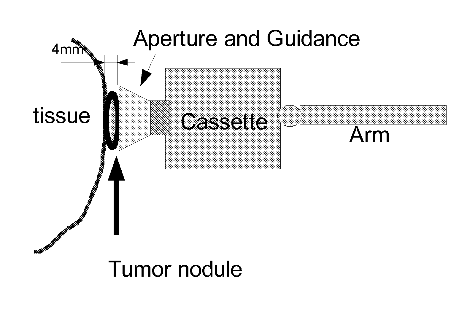
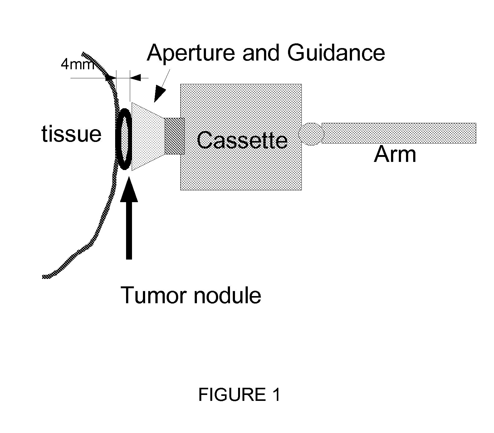
[0032]The preferred mode of invention proposes to first select an interchangeable irradiating capsule with a shutter as set forth below. Based on the depth and size of tissue to be treated, a radiation source will be selected for placement in the capsule and mounted on the robotic arm of the SRIORT. The arm would then be moved to the proper location for irradiation of the tissue, under direct visualization, with or without assistance from alternative imaging modalities or any combination of these.

[0033]Expanding on the above, the key invention components are:[0034]A radiation source[0035]A capsule / arm with an aperture opening to a cavity containing the radiation source with certain control electronics and devices designed to be connected to the surgical robot and inserted into the patient's body through the laparoscopic / surgical robotic incisions[0036]For a lesion, tumor, tissue, or organ, a mechanism for displaying pre-operative medical imaging, fused pre-operative medical imaging,...


PUM


Abstract

This invention proposes a robotic applicator device to be deployed internally to a patient having a capsule (also referred to as a cassette) and aperture with a means of alternately occluding and exposing a radioactive source through the aperture. The capsule and aperture will be integrated with a surgical robot to create a robotic IORT (intra-operative radiation therapy) applicator device as more fully described below. The capsule, radiation source, and IORT applicator arm would be integrated to enable a physician, physicist or technician to interactively internally view and select tissue for exposure to ionizing radiation in sufficient quantities to deliver therapeutic radiation doses to tissue. Via the robotic manipulation device, the physician and physicist would remotely apply radiation to not only the tissue to be exposed, but also control the length of time of the exposure. Control means would be added to identify and calculate margin and depth of tissue to be treated and the proper radiation source or radioactive isotope (which can be any particle emitter, including neutron, x-ray, alpha, beta or gamma emitter) to obtain the desired therapeutic effects. The invention enables stereotactical surgery and close confines radiation therapy adjacent to radiosensitive tissue.

Description

CONTINUATION DATA[0001]This application claims benefit of and as may be required is a continuation-in-part for any national stage, including the United States, of U.S. Provisional application 60 / 973,545 entitled “Direct visualization Robotic Intra-Operative Radiation Therapy Applicator Device” filed on Sep. 19, 2007 and from which benefit is claimed for PCT / US2008 / 077100 of the same name, and a U.S. provisional application 61 / 098,225 of the same name filed on Sep. 18, 2008, and a U.S. Provisional Application of this name filed on Sep. 18, 2009, all of which are incorporated by reference. This is a section 371 entry as well as a continuation-in-part of PCT / US2008 / 077100.FIELD OF INVENTION[0002]This invention relates to radiation cancer treatment by a mobile miniature capsule or cassette containing a radioactive source deployed internally to a patient which is robotically manipulated having an openable aperture to allow radiation emission to more precisely destroy tumors, especially t...

Claims


Application Information

Patent Timeline
Patent Type & Authority Patents(United States)
IPC IPC(8): A61F5/00
CPCA61N5/1001A61N5/1067A61N2005/1052A61N2005/1055A61N2005/1061
Inventor ROBERTS, WALTER A.SCHUMM, III, BROOKE
Owner SRIORT
Why Eureka
Try Eureka