Late potential detection

a technology of late potential and detection method, applied in the field of late potential detection, can solve the problems of benign or potentially life-threatening arrhythmias, high diagnostic value, high accuracy and resolution,

Active Publication Date: 2012-04-26
ST JUDE MEDICAL
View PDF6 Cites 8 Cited by
  • Summary
  • Abstract
  • Description
  • Claims
  • Application Information

AI Technical Summary

Benefits of technology

[0012]It is an objective to provide an improved late potential detection.
[0017]The determined parameter indicative of late potentials is of highly diagnostic value and can be used for risk assessment regarding arrhythmias, such as ventricular tachycardia and fibrillation. The parameter can be determined at high accuracy and resolution and without the intervention of a physician or without the need for physician visits.

Problems solved by technology

For instance, the heart may beat too fast or too slow, and may possibly beat regularly or irregularly.
Arrhythmias may be benign or potentially life-threatening.
This condition is a potentially life-threatening arrhythmia because it may lead to ventricular fibrillation (VF) and sudden death.
Ventricular fibrillation is always a medical emergency and can lead to death within minutes if left untreated.
The chaotic electrical impulses in the myocardium of the ventricle significantly reduce the effective blood pumping and may even cause a total stop of the pumping.
Even with the signal averaged ECG it can be very hard to detect any ventricular late potentials, which put high demands on the investigating physician.
The prior art solutions require the patient to visit the physician at the clinic in order to investigate whether the patient suffers from any ventricular late potentials, which of course is time and cost consuming for both the patient and the healthcare system.
Additionally, there is a significant risk of missing patients suffering a risk of arrhythmias using the prior art solution as it may happen that a patient can suffer from ventricular late potentials that come and go.
In such a case, there might be no ventricular late potentials detected during the visit at the clinic though the patient may still suffer from ventricular late potentials that occur at other time periods and therefore run a risk of arrhythmias.

Method used


Image

  • Late potential detection
  • Late potential detection
  • Late potential detection

Examples


Embodiment Construction

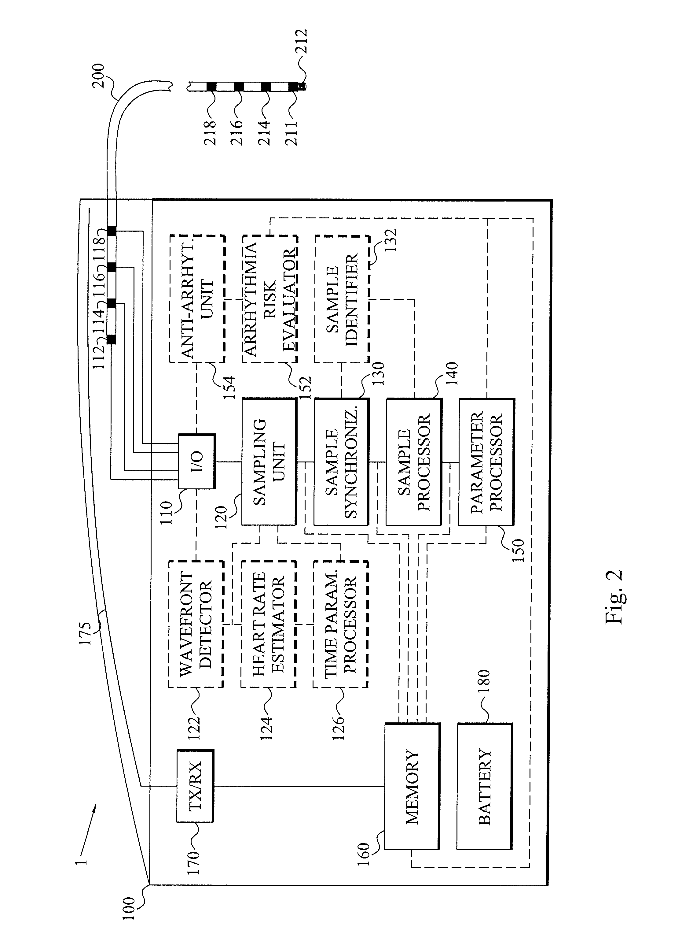
[0034]Throughout the drawings, the same reference numbers are used for similar or corresponding elements.

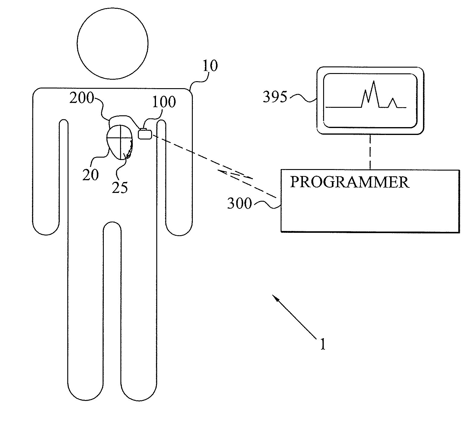
[0035]The present invention relates to an at least partly implantable system capable of automatically detecting late potentials for a patient, preferably mammalian patient and most preferably a human patient. The system comprises an implantable medical device arranged for sensing electrical activity of the heart of the patient and optionally capable of delivering electric treatment signals to the heart, for instance in the form of pacing pulses, cardioversion shocks and / or defibrillation shocks. The implantable medical device can therefore advantageously be a pacemaker, a defibrillator or a cardioverter.

[0036]The at least partly implantable system achieves significant advantages over the prior art late potential detections. For instance, no visits to a healthcare facility for the purpose of acquiring surface 12-lead electrocardiography (ECG) are required as the recording by the s...


PUM


Abstract

A late potential detecting system has an implantable medical device connected to at least one cardiac lead having implantable electrodes positioned at different sites of a ventricle myocardium. A sampling unit of the implantable medical device records electrogram samples for the different implantable electrodes to get different sample sets. The electrogram samples of the sample sets are time synchronized and magnitude potential representations of the potential data of the electrogram samples are determined. The magnitude potential representations of the time synchronized electrogram samples are then co-processed and used for determining a parameter that is indicative of any late potentials of the monitored ventricle.

Description

BACKGROUND OF THE INVENTION[0001]1. Field of the Invention[0002]The present invention generally relates to late potential detection, and in particular to a system comprising an implantable medical device for detecting late potentials of a cardiac ventricle.[0003]2. Description of the Prior Art[0004]Cardiac arrhythmia, sometimes denoted dysrhythmia, encompasses a large heterogeneous group of medical conditions in which there is abnormal electrical activity in the heart of a patient. For instance, the heart may beat too fast or too slow, and may possibly beat regularly or irregularly. Arrhythmias may be benign or potentially life-threatening. Ventricular tachycardia (VT) is an example of the latter and involves fast heart rhythms that originate in one of the ventricles of the heart. This condition is a potentially life-threatening arrhythmia because it may lead to ventricular fibrillation (VF) and sudden death. Ventricular fibrillation involves a multitude of micro-reentry circuits an...

Claims


Application Information

Patent Timeline
Patent Type & Authority Applications(United States)
IPC IPC(8): A61B5/0472A61B5/287A61B5/366A61B5/349
CPCA61B5/042A61N1/362A61B5/0452A61B5/686A61B5/0006A61B5/002A61B5/0031A61B5/7242A61B5/7275A61B2505/07G16H40/63G16H50/20A61B5/287A61B5/349
Inventor BLOMQVIST, ANDREAS
Owner ST JUDE MEDICAL
Why Eureka
Try Eureka