Method and apparatus for soft tissue retraction

a soft tissue retraction and soft tissue technology, applied in the field of soft tissue retraction, can solve problems such as operator fatigue, and achieve the effect of minimally invasive surgery

Active Publication Date: 2012-05-10
INVUITY
View PDF7 Cites 93 Cited by
  • Summary
  • Abstract
  • Description
  • Claims
  • Application Information

AI Technical Summary

Benefits of technology

[0006]The devices described below provide improved illumination for minimally invasive surgery and may include a retractor body with an improved retractor blade suitable for minimally invasive surgery. This illuminated retractor is useful in a wide array of surgical procedures performed in deep, restricted and / or awkward surgical fields including open and minimally invasive procedures.
[0008]The proximal projection may be configured for application of counter traction without the need for squeezing the retractor body which often leads to operator fatigue. The proximal projection may be weighted to balance the instrument as well as enabling the retractor to provide counter traction by itself. The configuration of the proximal projection further enables self-retraction by including a generally flat foot to prevent rolling and sliding of the retractor when it is providing self-retraction.
[0010]The retractor body may include a source of illumination and a portable source of energy to generate illumination. The proximal projection may be weighted to control the center of mass of the illuminated soft tissue retractor. The illumination waveguide may comprise a light input and a collar disposed thereover such that an air gap is maintained around the light input. The light input may have a generally cylindrical proximal portion transitioning into a rectangular distal portion, and the distal portion may be optically coupled with the illumination waveguide. The retractor may further comprise a shield that is disposed over the illumination waveguide which protects the illumination waveguide from damage caused by other surgical instruments. The shield may also prevent glare from shining back into a surgeon's eyes. The retractor blade may comprise a substantially rectangular portion and a flared distal tip. The retractor blade may form a drop angle with the retractor body, and the drop angle may range from 5 to 35 degrees. The retractor may further comprise a flat foot disposed on the proximal projection which facilitates self-retraction by preventing rolling and sliding of the retractor.
[0011]In another aspect of the present invention, a method of performing a minimally invasive thyroidectomy on a patient comprises providing an illuminated soft tissue retractor having a retractor blade, creating a minimally invasive surgical incision, and inserting the retractor blade into the incision. The soft tissue retractor also has a retractor body and an illumination waveguide. The retractor blade is coupled to the retractor body and the illumination waveguide is coupled to the retractor blade. The incision is preferably within 1 cm of the cricoids cartilage of the patient. The method also includes illuminating the surgical field with light extracted from the illumination waveguide. The light is directed laterally and distally from the illumination waveguide to illuminate the surgical field, and the light is transmitted through the illumination waveguide by total internal reflection. The soft tissue retractor retracts tissue thereby permitting a surgeon to perform a thyroidectomy or other procedure. The thyroidectomy may comprise using the soft tissue retractor to expose and dissect the patient's thyroid. The retractor blade may be used to bluntly dissect tissue, or the retractor blade may also be used to counter retract tissue. Counter retracting tissue may comprise providing a proximal projection disposed adjacent a proximal end of the retractor body such that the counter retraction may be applied without squeezing the retractor body. Creating the minimally invasive surgical incision may comprise making an incision no longer than 3 to 4 cm long. The method may also comprise protecting the waveguide illuminator by providing a shield that is disposed adjacent the waveguide illuminator. The shield may also block glare by preventing the glare from shining back into an operator's eyes.

Problems solved by technology

The proximal projection may be configured for application of counter traction without the need for squeezing the retractor body which often leads to operator fatigue.

Method used


Image

  • Method and apparatus for soft tissue retraction
  • Method and apparatus for soft tissue retraction
  • Method and apparatus for soft tissue retraction

Examples


Embodiment Construction

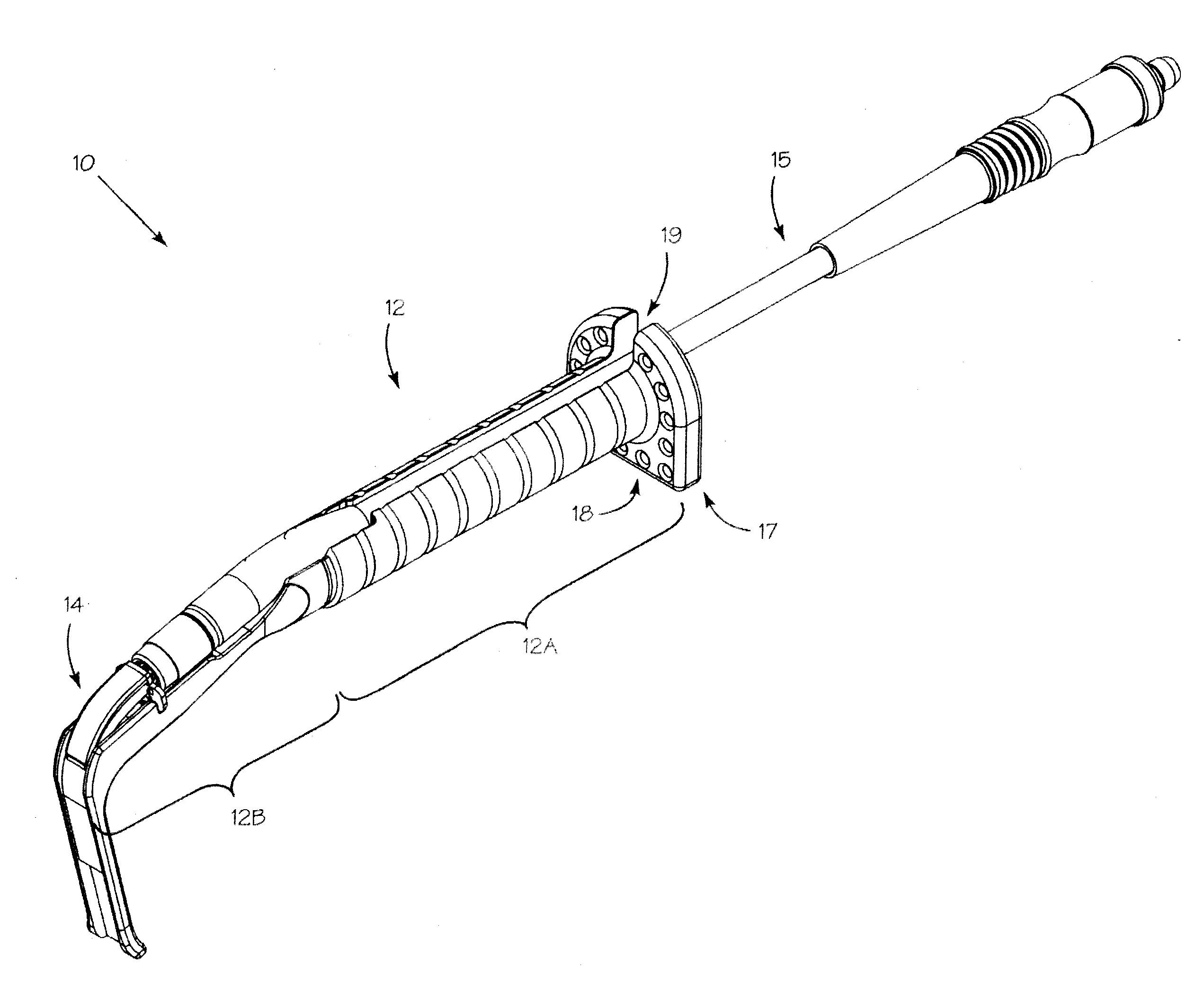
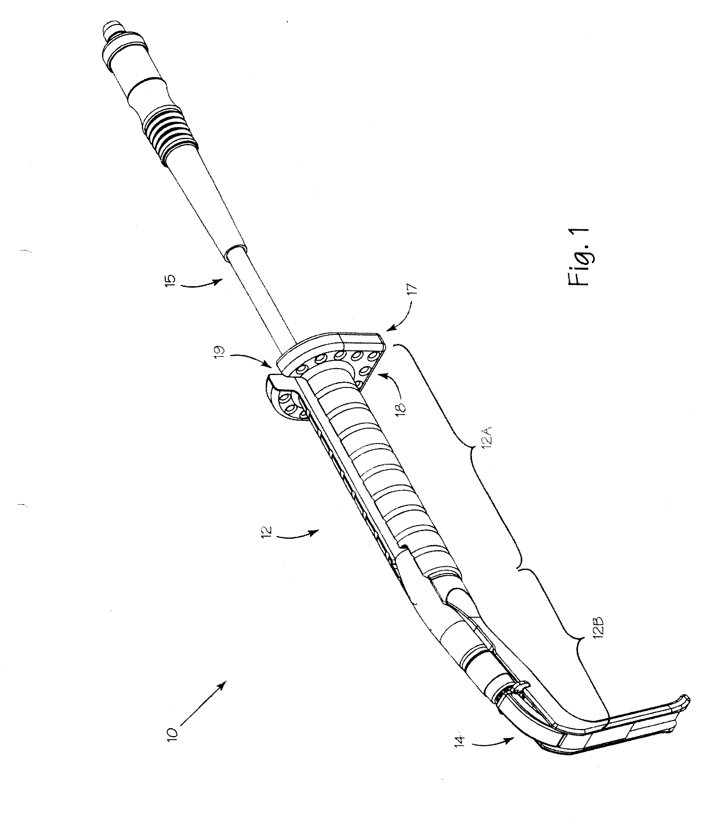
[0022]Referring to FIG. 1, illuminated soft tissue retractor 10 includes retractor assembly 12, illumination waveguide assembly 14 and illumination assembly 15. Proximal projection 17 extends generally perpendicular from retractor body 12A. Retractor blade 12B is coupled with a distal portion of the retractor body 12A and may include a proximal portion that is generally lies in the same plane as the retractor body 12A, and a distal portion which is transverse thereto. In some embodiments, the distal portion of the retractor blade is orthogonal to the proximal portion of the retractor blade, although other angles may be used. Proximal projection 17 optimizes application of counter traction without the need for squeezing retractor body 12A which often leads to fatigue. Proximal projection 17 may be weighted to balance the instrument as well as enabling the retractor to provide counter traction by itself Proximal projection 17 may be formed of heavier material than retractor body 12A o...


PUM


Abstract

A soft tissue retractor includes a retractor body and a proximal projection extends generally perpendicular to the proximal end of the retractor body. An illumination waveguide engages a slot in the retractor blade to provide illumination to a surgical site while maintaining total internal reflection in the waveguide. An illumination input is formed into the proximal end of the illumination waveguide for conducting light from a source to the illumination waveguide. The proximal projection is configured for application of counter traction without the need for squeezing the retractor body. The proximal projection may be weighted to balance the soft tissue retractor as well as enabling the retractor to provide counter traction by itself. The configuration of the proximal projection further enables self-retraction by including a flat foot to prevent rolling and sliding of the retractor when it is providing self-retraction.

Description

CROSS-REFERENCE[0001]The present application is a non-provisional of, and claims the benefit of U.S. Provisional Patent Application No. 61 / 391,512 (Attorney Docket No. 40556-717.101, formerly 028638-001500US) filed Oct. 8, 2010; the entire contents of which is incorporated herein by reference.BACKGROUND OF THE INVENTION[0002]The present invention relates generally to the field of soft tissue retraction for minimally invasive surgery and more specifically to methods and illumination systems with integrated retractors for minimally invasive soft tissue retraction.[0003]In various surgical procedures, illumination of the surgical field is typically achieved through the use of overhead lighting, headlamps and surgical microscopes. There are scenarios in which these illumination sources provide lighting that is either poor in quality or poorly directed. Light from an overhead source is fixed and often blocked by the surgeon or others in the operating room. Surgical microscopes are little...

Claims


Application Information

Patent Timeline
Patent Type & Authority Applications(United States)
IPC IPC(8): A61B1/06A61B1/32
CPCA61B1/0676A61B19/5202A61B17/02A61B1/32A61B90/30A61B1/07A61B17/0218
Inventor VAYSER, ALEXCOVEY, EDWARD A.BREDENKAMP, JAMES K.STOCK, LELAND A.
Owner INVUITY
Why Eureka
Try Eureka