Visual tracking and annotaton of clinically important anatomical landmarks for surgical interventions

a technology of anatomical landmarks and visual tracking, applied in the field of systems and methods of acquiring and displaying information, can solve the problems of time-consuming, add to the cognitive load of the surgeon, and the anatomy may deform

Active Publication Date: 2012-09-06
THE JOHN HOPKINS UNIV SCHOOL OF MEDICINE
View PDF27 Cites 142 Cited by
  • Summary
  • Abstract
  • Description
  • Claims
  • Application Information

AI Technical Summary

Problems solved by technology

If the surgeon does not recall or is not sure about the location or the content of a prior diagnostic image associated with a particular area, the diagnostic device might have to be reintroduced to the operating field, which is time consuming.
This is difficult when the treatment is inside the target tissue, or does not alter the tissue's appearance.
This adds to cognitive load on the surgeon in an already challenging minimally invasive procedure.
Furthermore, the anatomy may deform, or change color naturally, or from the intervention itself, which adds to the difficulty of tracking the relevant landmarks and associated intraoperative information.
The resulting data is sparsely located, requiring the surgeon to track multiple scans or images with the corresponding anatomical locations in the microscope view, which adds significant cognitive load to the already challenging surgical task.
This can become more difficult with altered anatomy due to surgical manipulation, bleeding, bio-markers, swelling as well as inherent changes in the field of view, lighting methods and / or directions and intraocular fluid conditions.

Method used


Image

  • Visual tracking and annotaton of clinically important anatomical landmarks for surgical interventions
  • Visual tracking and annotaton of clinically important anatomical landmarks for surgical interventions
  • Visual tracking and annotaton of clinically important anatomical landmarks for surgical interventions

Examples


examples

[0039]The following are a couple examples of the use of a visual tracking and annotation system according to some embodiments of the current invention. These examples are provided for illustration and are not intended to define the broad scope of the current invention.

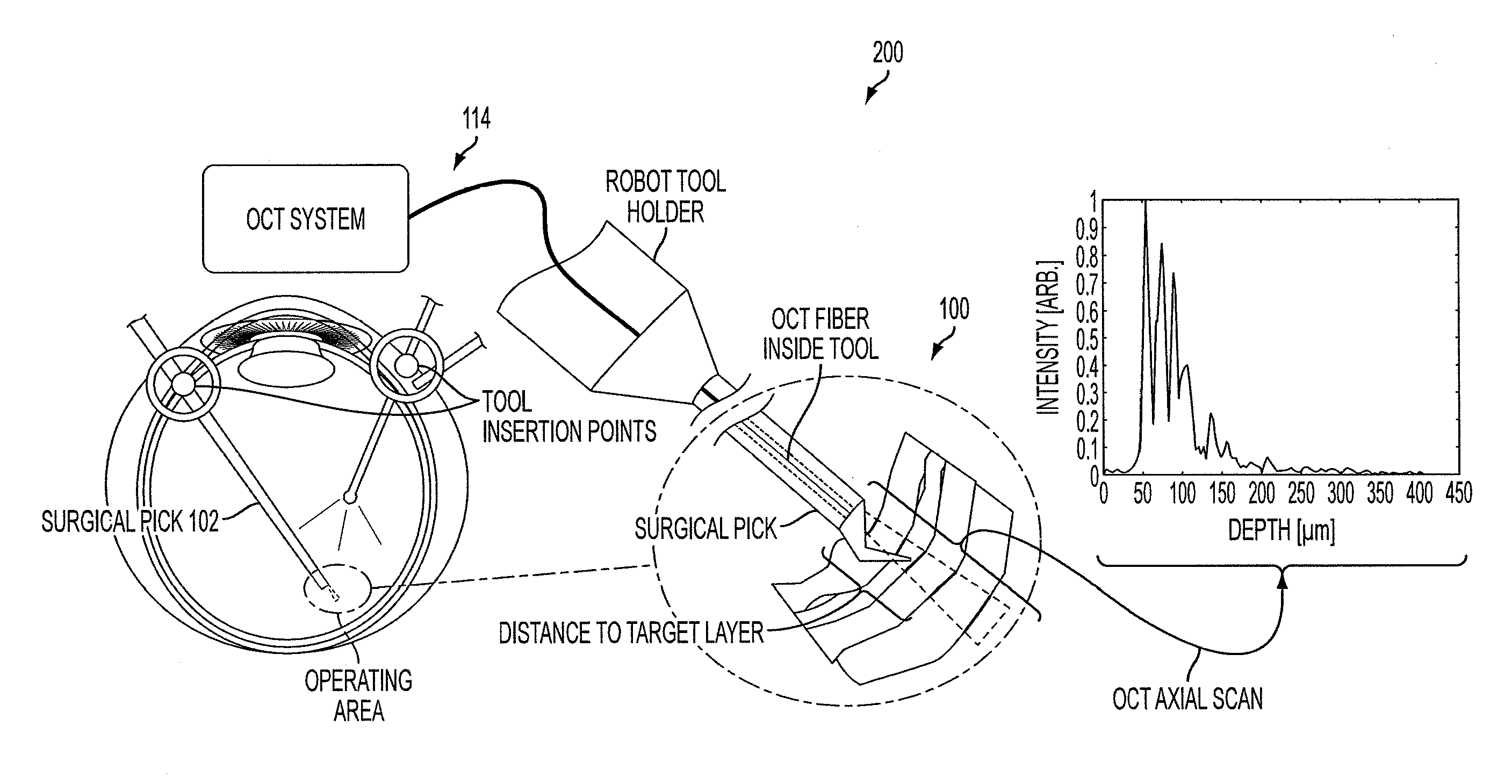
[0040]VitroRetinal Surgery

[0041]The surgeon brings the OCT probe near an area of interest

[0042]While a foot pedal is pressed (or voice command actuation) the surgeon sweeps the probe across an area of interest

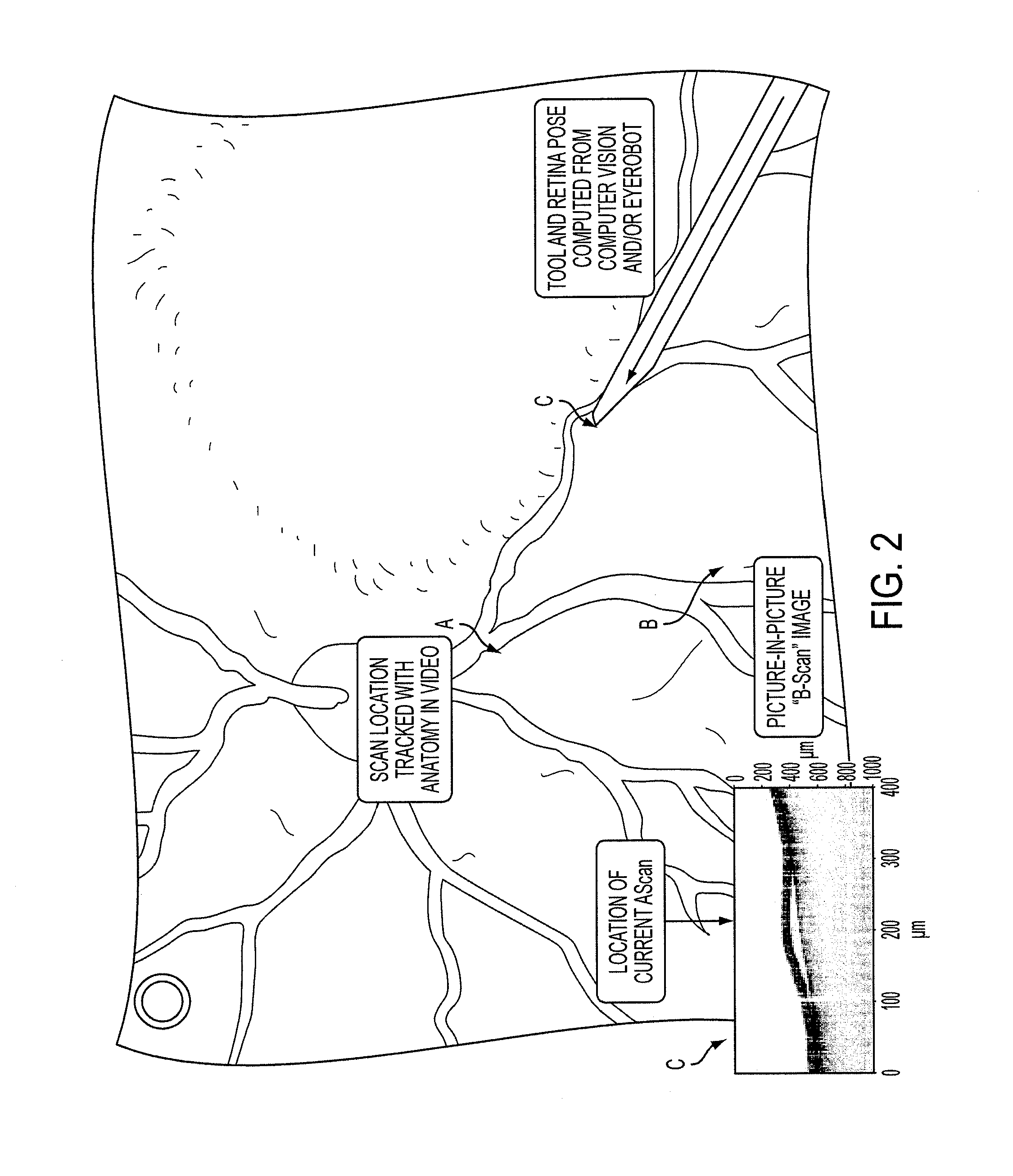
[0043]The location of the scan is tracked relative to the tissue and projected on the video feed via visual and / or external tool tracking. The time synchronized sensor data stream is associated with this path.

[0044]From this point on the tagged anatomical area is tracked and annotated accordingly

[0045]The surgeon may choose to create another scan or investigate reexamine the existing ones.

[0046]The surgeon can request vocally to review “scan number 3” or hover over it with the surgical instrument. The


PUM


Abstract

A visual tracking and annotation system for surgical intervention includes an image acquisition and display system arranged to obtain image streams of a surgical region of interest and of a surgical instrument proximate the surgical region of interest and to display acquired images to a user; a tracking system configured to track the surgical instrument relative to the surgical region of interest; a data storage system in communication with the image acquisition and display system and the tracking system; and a data processing system in communication with the data storage system, the image acquisition and display system and the tracking system. The data processing system is configured to annotate images displayed to the user in response to an input signal from the user.

Description

CROSS-REFERENCE OF RELATED APPLICATION[0001]This application claims priority to U.S. Provisional Application No. 61 / 256,531 filed Oct. 30, 2009, the entire contents of which are hereby incorporated by reference.[0002]This invention was made with Government support of Grant No. 1R01 EB 007969-01, awarded by the Department of Health and Human Services, NIH; and Grant No. EEC-9731478, awarded by the NSF. The U.S. Government has certain-rights in this invention.BACKGROUND[0003]1. Field of Invention[0004]The field of the currently claimed embodiments of this invention relates to systems and methods of acquiring and displaying information during surgical procedures, and more particularly to systems and methods of acquiring and displaying information that include visual tracking of surgical instruments and annotation of information displayed.[0005]2. Discussion of Related Art[0006]Currently surgeons use a number of intraoperative diagnostic and surgical and / or treatment devices for operati...

Claims


Application Information

Patent Timeline
Patent Type & Authority Applications(United States)
IPC IPC(8): A61B6/02A61B8/13A61B5/103A61B6/00
CPCA61B5/0066A61F9/007A61B3/102A61B3/1225A61B2019/5276A61B8/5261A61B2019/5231A61B3/13G02B21/0012A61B90/37A61B2034/2065A61B2090/3614A61B2090/3735A61B5/743A61B5/7475A61B5/0084A61B5/061A61B5/1076A61B2560/0295G06T7/33G16H20/40G16H30/40A61B2090/373A61B2090/378G06F40/169
Inventor BALICKI, MARCIN A.TAYLOR, RUSSELL H.HAGER, GREGORY D.GEHLBACH, PETER L.HANDA, JAMESKUMAR, RAJESH
Owner THE JOHN HOPKINS UNIV SCHOOL OF MEDICINE
Why Eureka
Try Eureka