
Method and magnetic resonance tomography system to generate magnetic resonance image data of an examination subject

Active Publication Date: 2012-09-13
SIEMENS HEALTHCARE GMBH
View PDF4 Cites 17 Cited by
  • Summary
  • Abstract
  • Description
  • Claims
  • Application Information

AI Technical Summary

Benefits of technology

[0009]An object of the present invention is to provide a method as well as a suitable magnetic resonance tomography system that avoid the problem described above, even in data acquisition procedures in which the table is moved during the magnetic resonance measurement.

Problems solved by technology

This can significantly hinder the assessment of the images by the radiologist, and increases the risk of false positive findings and also hinders the evaluation of the images with computer-assisted methods.
The sensitivity of the local coil therefore changes during the measurement, and the known methods for correction of the unwanted intensity variations can no longer be used.

Method used


Image

  • Method and magnetic resonance tomography system to generate magnetic resonance image data of an examination subject
  • Method and magnetic resonance tomography system to generate magnetic resonance image data of an examination subject
  • Method and magnetic resonance tomography system to generate magnetic resonance image data of an examination subject

Examples


Embodiment Construction

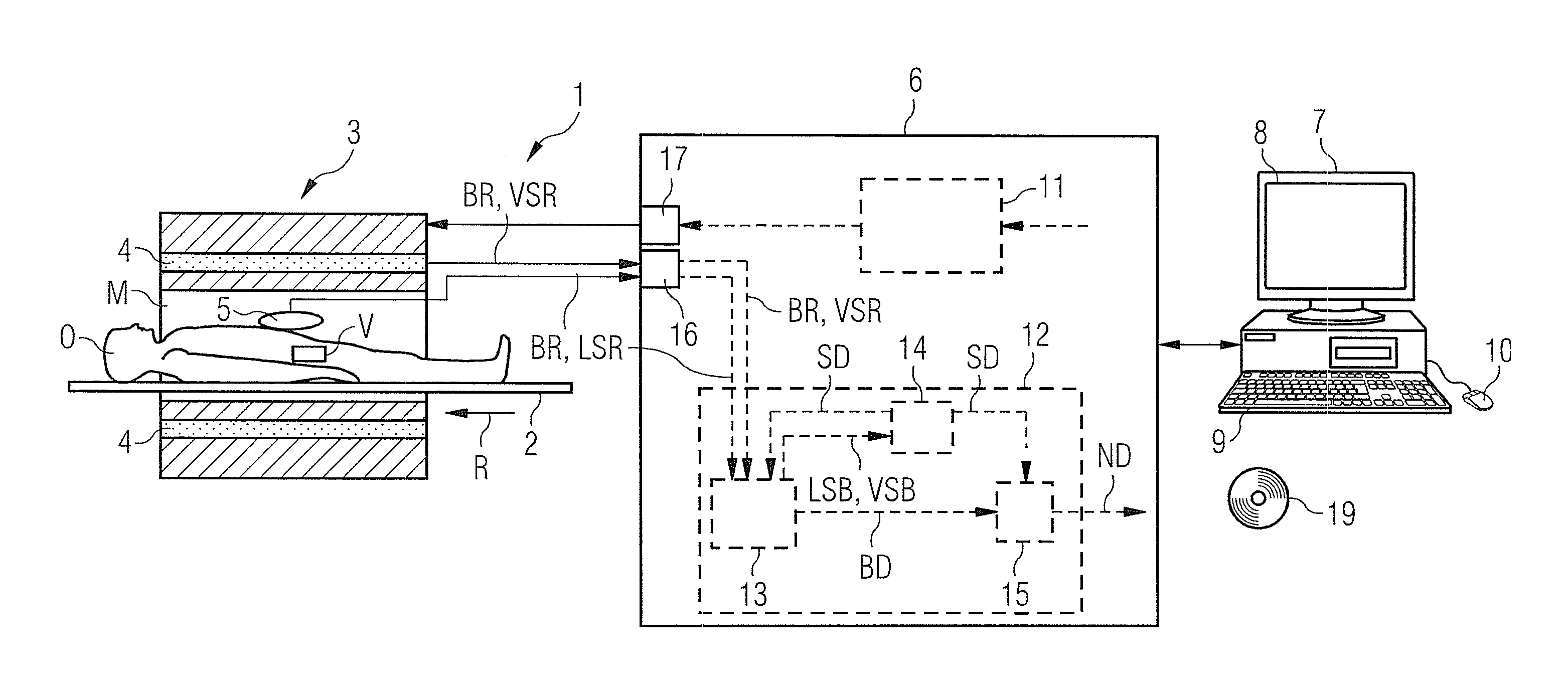
[0039]An exemplary embodiment of a magnetic resonance tomography system 1 according to the invention is schematically presented in FIG. 1. The magnetic resonance tomography system 1 essentially includes a magnetic resonance scanner (data acquisition unit) 3 with which the basic magnetic field necessary for the magnetic resonance measurement is generated in a measurement space M. A table 2 on which a patient or examination subject O can be positioned is located in the measurement space M (also called a patient tunnel). As is typical, the magnetic resonance scanner 3 has a volume coil 4 as a transmission antenna system. Moreover, multiple gradient coils are located in the scanner 3 in order to respectively apply the desired magnetic field gradients in the three spatial directions. For clarity, however, these components are not shown in FIG. 1.

[0040]The magnetic resonance tomography system 1 also has a control device 6 with which the scanner 3 is controlled and magnetic resonance data,...


PUM


Abstract

A method and magnetic resonance tomography system to generate magnetic resonance image data of an examination subject, raw imaging data are acquired from multiple slices of a predetermined volume region of the examination subject using local coils during a table feed in the magnetic resonance scanner. Image data of the slices are reconstructed on the basis of the raw imaging data. A normalization of the image data is subsequently implemented on the basis of measured coil sensitivity data of the local coils that are used.

Description

BACKGROUND OF THE INVENTION[0001]1. Field of the Invention[0002]The invention concerns a method to generate magnetic resonance image data of an examination subject by means of a magnetic resonance tomography system, in which method raw imaging data composed of multiple slices of a predetermined volume region of the examination subject are acquired using local coils during a table feed into the magnetic resonance scanner during a magnetic resonance measurement (data acquisition), and based on this raw imaging data, image data of the slices are then reconstructed. Moreover, the invention concerns a magnetic resonance tomography system with a magnetic resonance scanner and a control device designed to control the magnetic resonance tomography system for a magnetic resonance measurement so that raw imaging data composed of multiple slices of a predetermined volume region of an examination subject are acquired using local coils during a table feed into the magnetic resonance scanner, and...

Claims


Application Information

Patent Timeline
IPC IPC(8): G01R33/44
CPCG01R33/246G01R33/5659G01R33/56383
Inventor STEMMER, ALTO
Owner SIEMENS HEALTHCARE GMBH
Learn More