Biomarkers of musculoskeletal disease
a biomarker and musculoskeletal technology, applied in the field of musculoskeletal disease biomarkers, can solve the problems of slowing the overall rate of deamidation in joint tissues, excessive production or degradation, and serious and irreversible damage to cartilage fibrillar meshwork
- Summary
- Abstract
- Description
- Claims
- Application Information
AI Technical Summary
Problems solved by technology
Method used
Image
Examples
example 1
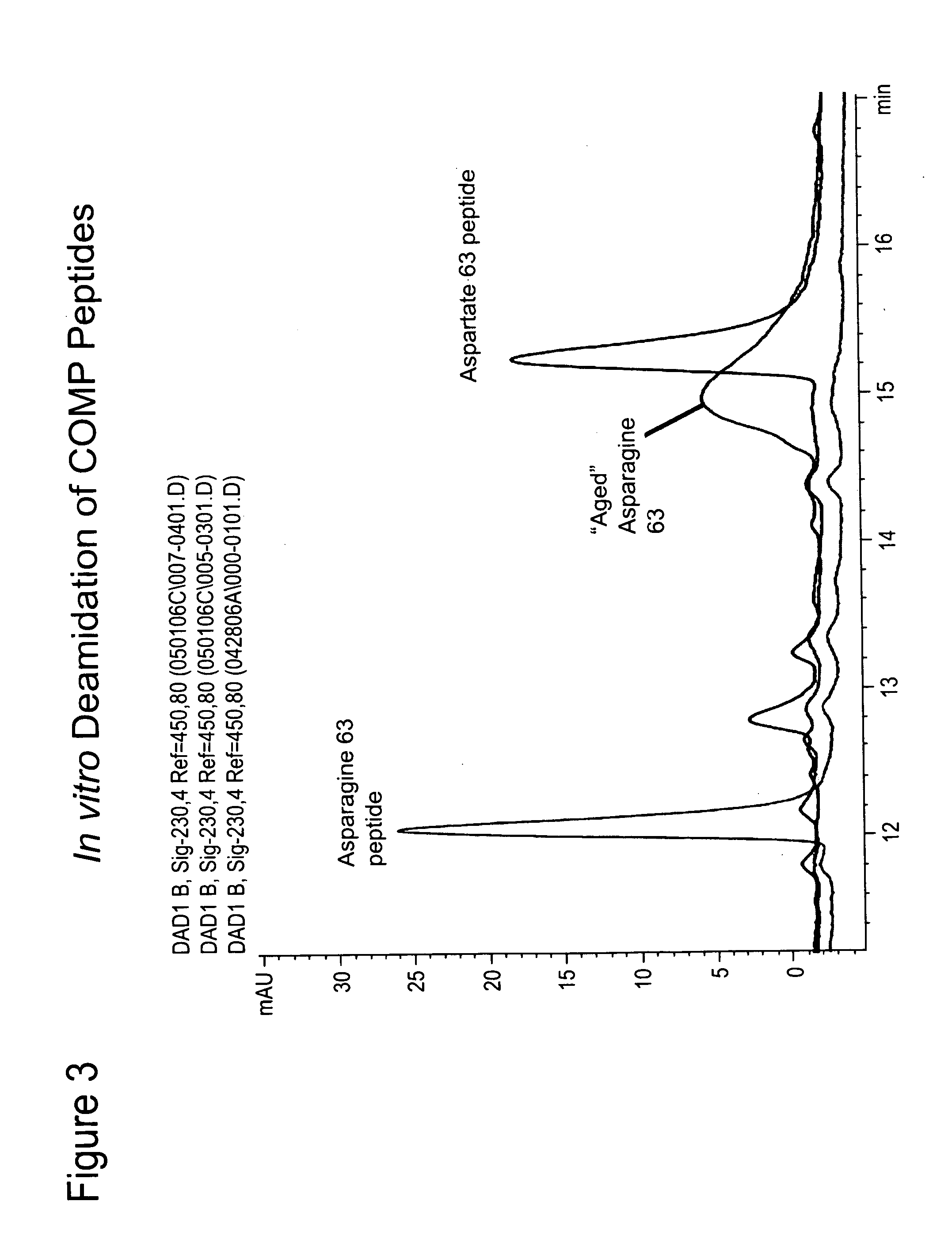
[0074]As described below, peptides have been identified within COMP that are predicted to undergo deamidation. Monoclonal antibodies recognizing the deamidated forms have been developed and their specificity for biomarker studies validated. In the course of this work, a method of aging native COMP peptides to induce deamidation, verified by HPLC analysis, has been developed.
[0075]The native Asn containing COMP (DUK006A) peptide (see Example 2) was incubated at room temperature for 72 hours leading to a rightward shift in the HPLC trace resulting in its overlap with the migration of the corresponding deamidated COMP peptide (FIG. 3). It is possible to differentiate the native (FIG. 4A) and deamidated (FIG. 4B) epitopes by mass spectroscopy and the presence of both in a mixture (FIG. 4C) (Doyle et al, Ann. N.Y. Acad. Sci. 1050:1-9 (2005)). A similar procedure can be used for identifying hot spots of deamidation in purified fragments of cartilage ECM proteins.
example 2
[0076]Described below is the development and validation of monoclonal antibodies to deamidated and / or native residues in COMP. Antibodies described are suitable for use in the detection of biomarkers.
[0077]Putative deamidation hotspots within COMP were determined using an algorithm developed by Robinson and Robinson (Molecular Clocks: Deamidation of Asparaginyl and Glutaminyl Residues in Peptides and Proteins, Cave Junction, Oreg., Althouse Press (2004); found at www.deamidation.org). This algorithm uses an investigator inputted crystal structure and amino acid composition of a protein to predict Asn residues susceptible to deamidation. The COMP protein structure files were retrieved from http: / / www.rcsb.org / in the form of the PDB files: 1 fbm.pdb and 1 vdf.pdb. Both files are for Rattus norvegicus COMP as the crystal structure for human COMP was not available. Using the rat sequence and structure, two different predicted hotspots were identified, Asn41 and Asn63. These sites corre...
example 3
[0094]Example 2 details the development of antibodies that are specific for a predicted deamidated epitope in COMP. Their specificity and the ability to recognize COMP in cartilage extracts is shown by western blot. Two questions are addressed by the studies described below:
[0095]1. Can an ELISA for aged deamidated COMP be developed?
[0096]2. Can the epitope be detected in body fluids?
[0097]One of the biggest problems faced when developing ELISA protocols to the epitopes described herein was what to use as a standard. The age-related changes are likely to be only present in a small subset of the total proteins making the use of native proteins an unlikely source. Generation of recombinant proteins for each of the epitopes, however, would be both time consuming and expensive.
[0098]A novel competition ELISA has been developed that uses the peptide sequence used originally to generate the antibodies, coupled to BSA (FIG. 10). With one of the monoclonal antibodies (6-1A12) to a COMP deam...
PUM
| Property | Measurement | Unit |
|---|---|---|
| pH | aaaaa | aaaaa |
| concentration | aaaaa | aaaaa |
| concentration | aaaaa | aaaaa |
Abstract
Description
Claims
Application Information
 - R&D
- Intellectual Property
- Life Sciences
- Materials
- Tech Scout
- Unparalleled Data Quality
- Higher Quality Content
- 60% Fewer Hallucinations
Browse by: Latest US Patents, China's latest patents, Technical Efficacy Thesaurus, Application Domain, Technology Topic, Popular Technical Reports.
© 2025 PatSnap. All rights reserved.Legal|Privacy policy|Modern Slavery Act Transparency Statement|Sitemap|About US| Contact US: help@patsnap.com


