
Radiology verification system and method

a radiology and report verification technology, applied in the field of system and method verification of radiology reports, can solve the problems of time constraints for radiologists to produce accurate yet complete readings, and achieve the effects of reducing radiologist time and effort, increasing accuracy and completeness, and reducing errors

Inactive Publication Date: 2012-12-13
LIPMAN BRIAN T
View PDF19 Cites 11 Cited by
  • Summary
  • Abstract
  • Description
  • Claims
  • Application Information

AI Technical Summary

Benefits of technology

[0006]With use of the techniques described herein, the accuracy and completeness of the produced radiology report may significantly increase. Further, implementation of the verification techniques into existing radiology imaging GUIs and radiology work flows enables increased accuracy and completeness with minimal radiologist time and effort.

Problems solved by technology

Radiologists are often under time constraints to produce an accurate yet complete reading of the radiology images, particularly in emergency or time-sensitive settings.

Method used


Image

  • Radiology verification system and method
  • Radiology verification system and method
  • Radiology verification system and method

Examples


Embodiment Construction

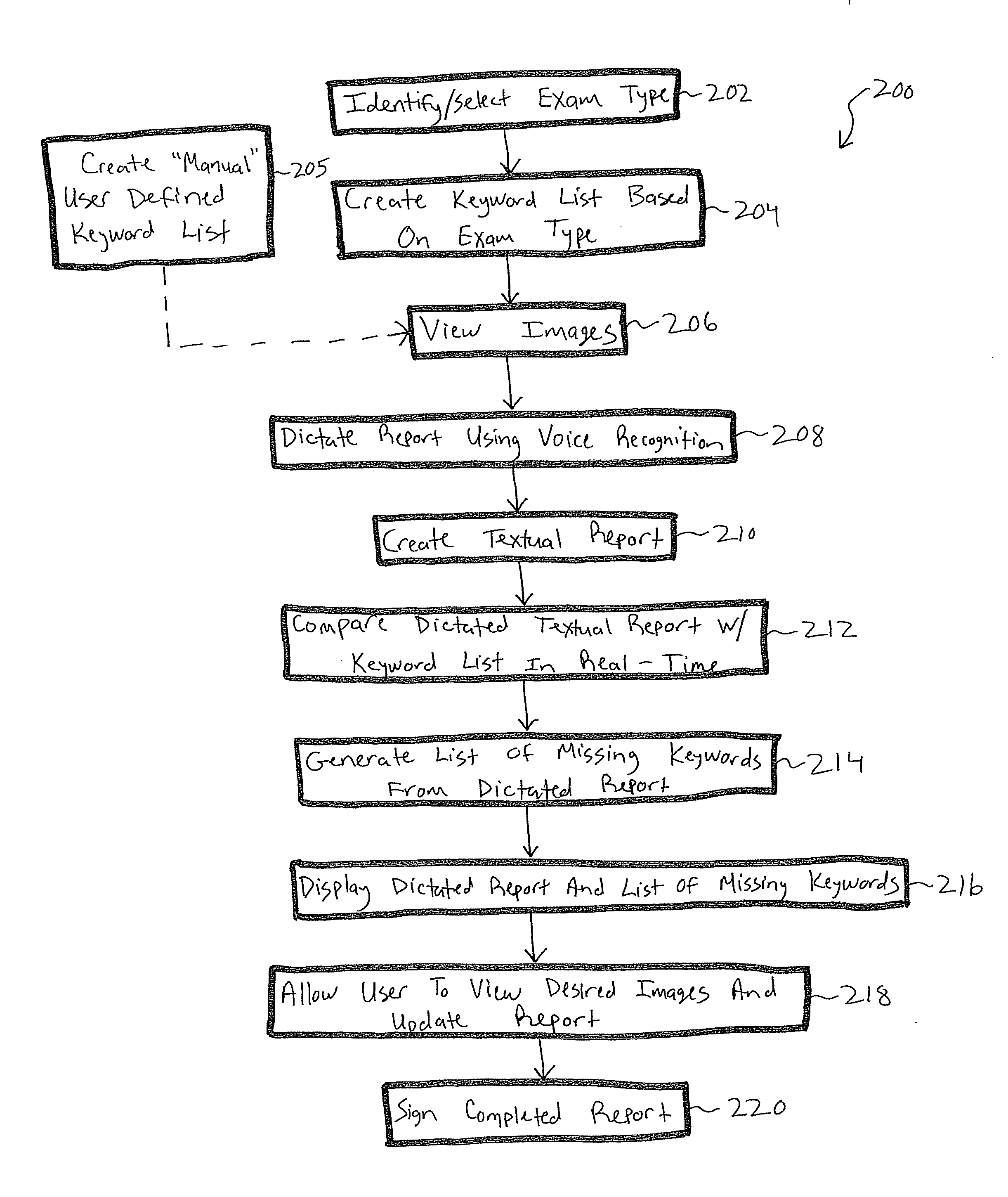
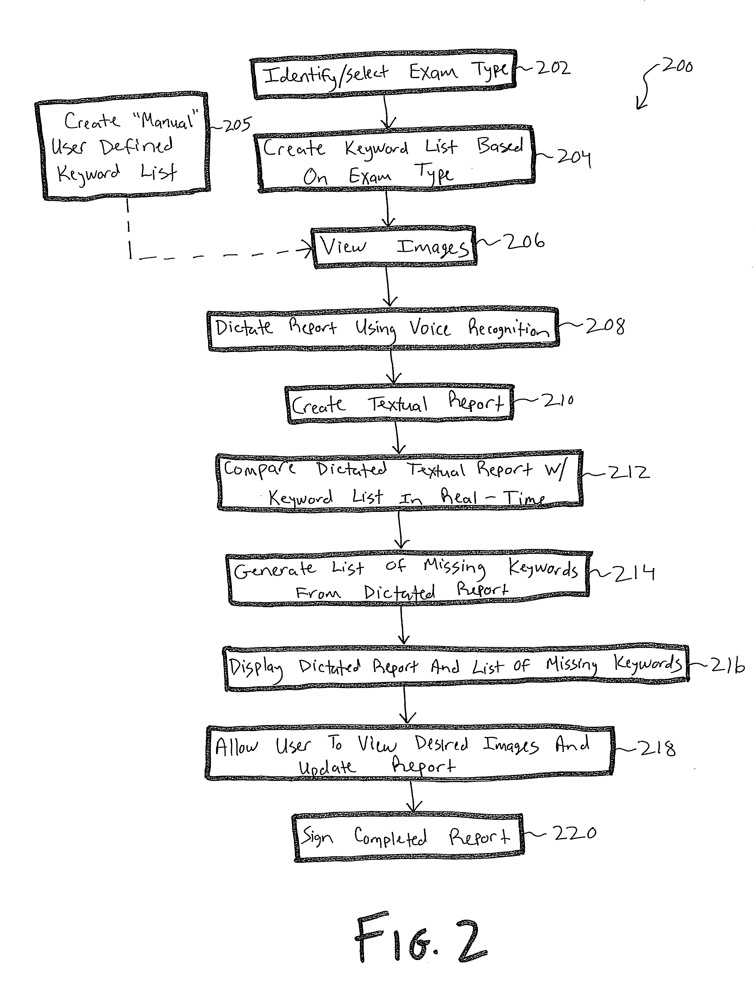
[0009]One embodiment of the present invention provides a series of verification techniques and methods to ensure that a radiology report contains a complete description. These verification techniques may be run against the radiology report during and after the read by the radiologist to ensure that the report does not lack a description for important anatomical or physiological features. As one example, when a radiologist dictates a radiological report, there are key anatomic structures (such as organs, bones, tissue) that he or she should comment on. The verification techniques described herein check for mention of these structures and prompt the radiologist as necessary to provide details on the structures. The various prompts and suggestions presented to the radiologist may be customized based on radiologist preferences, the type of radiological scan procedure, or the type of medical procedure or suspected medical diagnosis.

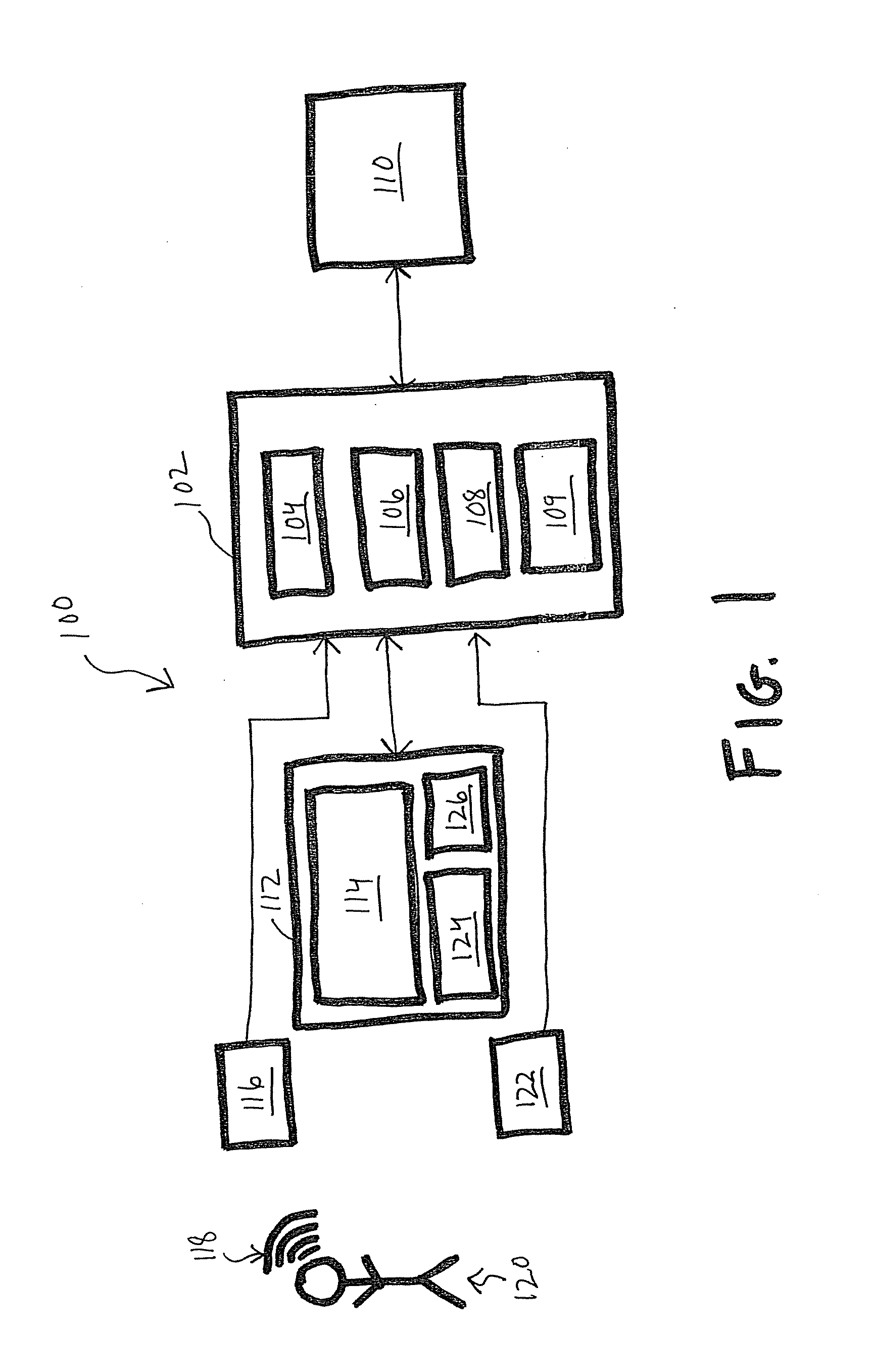
[0010]FIG. 1 provides an overview of a computing system ...


PUM


Abstract

A system and method of radiology verification is provided. The verification may be implemented as a standalone software utility, as part of a radiology imaging graphical user interface, or within a more complex computing system configured for generating radiology reports.

Description

FIELD OF THE INVENTION[0001]The present invention relates generally to systems and methods for verifying radiology reports. More particularly, the invention relates to a system and method of using voice recognition software to identify missing keywords in a radiology report.BACKGROUND OF THE INVENTION[0002]Most radiologists review digitized radiology images with use of a computer display, as electronic versions of the radiology images are rendered in a software graphical user interface (GUI) operating on a computer system. Radiologists often use voice recognition software that interfaces with the graphical user interface to dictate interpretations of the radiology images in an expeditious manner. The use of a voice recognition dictation system allows observations of the radiology images to be quickly dictated by the radiologist into the computer system, as a written report is immediately generated from the dictation for use by the medical facility or physician treating the patient.[...

Claims


Application Information

Patent Timeline
IPC IPC(8): G10L15/26
CPCG10L15/22G06Q50/24G06Q10/10G16H15/00G16H30/00
Inventor LIPMAN, BRIAN T.
Owner LIPMAN BRIAN T
Learn More