

Ophthalmologic apparatus, ophthalmologic system, controlling method for ophthalmologic apparatus, and program for the controlling method
a technology of ophthalmologic equipment and control method, which is applied in the field of ophthalmologic equipment, ophthalmologic system, and controlling method of ophthalmologic equipment, can solve the problem of restricting the speed of acquiring one oct tomographic image, and achieve the effect of correcting the curvature of the retina
- Summary
- Abstract
- Description
- Claims
- Application Information
AI Technical Summary
Benefits of technology
Problems solved by technology
Method used
Image

Examples
first embodiment
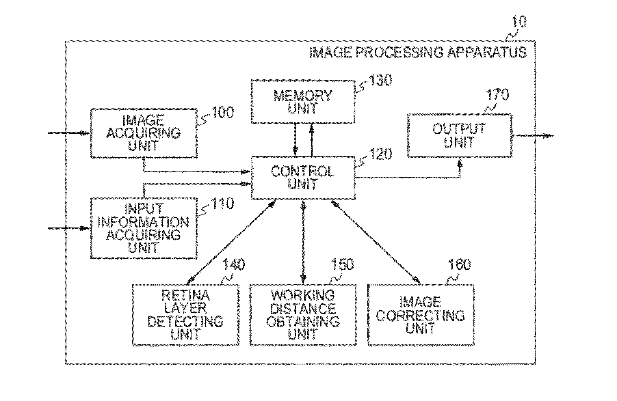
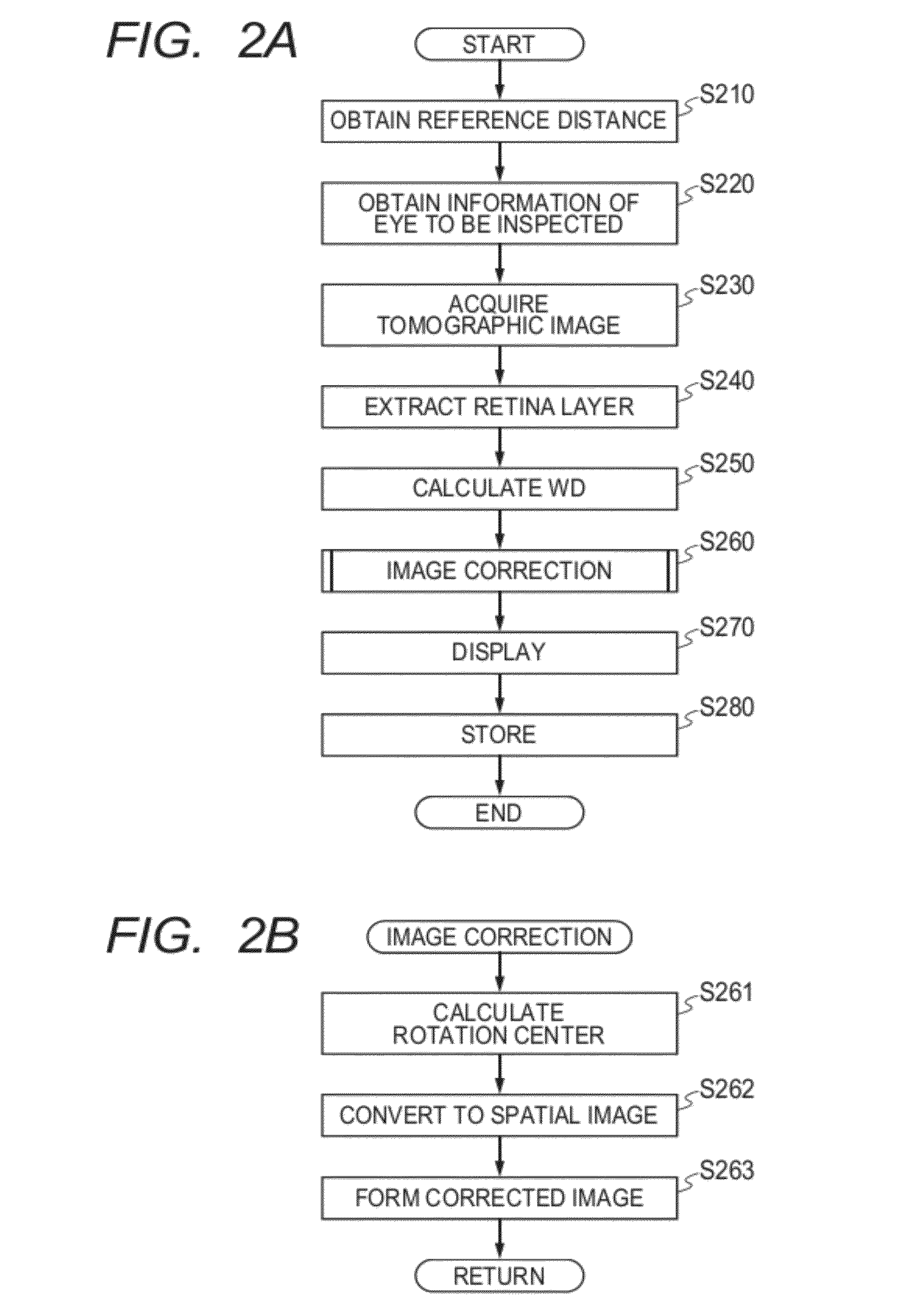
[0034]In a first embodiment of the present invention, when performing quantitative measurement of curvature of retina of a diseased eye such as a myopic eye that is known to have a large curvature, variation of curvature of retina of the acquired tomographic image is corrected by using a working distance (WD) obtained when the image is acquired so that a correct curvature is measured. More specifically, an axial length of an eye to be inspected is obtained, the WD is calculated from a coherence gate position when the image is acquired and a retina position in an acquired tomographic image, and the acquired tomographic image is corrected based on this WD value. With this correction, a correct curvature can be measured, and comparison among eyes to be inspected or evaluation of the variation with time can be performed.
[0035]In other words, a distance from a reference point to an objective lens is obtained, and the axial length of the eye to be inspected, the coherence gate position wh...
second embodiment
[0105]In a second embodiment of the present invention, when acquiring the working distance when the image is acquired by the method described above in the first embodiment, the acquired working distance is compared with the working distance of the image that has been acquired before for the same eye to be inspected, and a difference between the working distances is displayed so as to assist the operator to acquire an image at the same working distance. The first embodiment describes that it is possible to correct the image to have the same curvature even if the images are acquired at different working distances. However, in general, image quality of the corrected image is deteriorated from that of the original tomographic image. Therefore, if the image is acquired at a working distance as close as possible to the working distance when the image was acquired last time, it is possible to compare the retina state between images without correction.
[0106]A functional configuration of the...
third embodiment
[0122]In the second embodiment, description is given of a method in which the image is acquired at the same working distance as that when the image was acquired last time, so as to enable comparison of the retina including the curvature between the images without the correction, too. However, in a myopic eye, when inspecting an eye in which progress of posterior staphyloma or the like is observed, there may be a case where elongation of the axial length is observed in a progress observation. In this case, if the working distance is equalized (supposing that there is no change with time in the anterior eye part), the rotation center for scanning of the measuring light can be positioned at a position of the same positional relationship from the anterior eye part. However, the distance from the rotation center to the retina is changed when the axial length is elongated, and hence it is difficult to compare between the obtained images even if the coherence gate position is adjusted.
[012...
PUM
 Abstract
Description
Claims
Application Information