
Interactive analytics of digital histology slides

an analysis and slides technology, applied in the field of digital pathology, can solve the problems of inability to perform interactively, large size of the roi, and inability to perform histopathological analysis in a real-time manner, and achieve the effect of minimizing perceptible delay

Inactive Publication Date: 2013-02-07
NEC CORP
View PDF8 Cites 4 Cited by
  • Summary
  • Abstract
  • Description
  • Claims
  • Application Information

AI Technical Summary

Benefits of technology

The patent describes a method and system for analyzing images in real-time with user interaction. The interface layer receives the selection of an image and requests analysis, which is then transferred to the interpretation layer. The interpretation layer divides the selected region of the image into smaller parts for parallel computation, resulting in faster analysis and minimized delay between receiving the request and results. The analysis is performed by execution nodes with processors, which receive parts of the selection and perform the requested analysis on them. The system includes a shared memory for storing full images and analysis results, and an execution controller for optimizing analysis performance by dividing the selected region of the image into sub-sections. The technical effects of the patent include faster and more efficient image analysis with user interaction.

Problems solved by technology

Image analysis normally operates on a portion of the image, called the region of interest (ROI), but the size of the ROI is often constrained by the complexity of the analysis and the available computing resources.
Analytics on histopathological images, including image processing, image analysis, and machine learning, is frequently computationally intensive and cannot be performed in an interactive way.
These systems do not have the capability of executing full analysis of tissues.
As a result, analysis on such systems is not scalable, and demanding functions take too long for interactive execution.
Furthermore, existing distributed computing systems are inadequate to address the needs of digital pathology, because the computation and communication demands may overwhelm even powerful distributed systems.
Images of histology slides can be, for example, several gigabytes in size, such that it is usually infeasible to transfer such images back and forth between client and server.
Moreover, the computations involved in modern analytics can be very intensive, particularly if performed on the entire image.
As such, existing cloud servers are not optimized to handle digital pathology services.

Method used


Image

  • Interactive analytics of digital histology slides
  • Interactive analytics of digital histology slides
  • Interactive analytics of digital histology slides

Examples


Embodiment Construction

[0017]The present principles provide a multi-layer system that allows for distributed processing of analytical tasks, allowing users to perform analysis in real-time with a high degree of responsiveness. Processing is separated into a user interface layer that permits a user to interactively view and direct analysis, and an interpretation layer that distributes computation-heavy analysis to a back-end server or servers that have greater computational power than the interface layer. By focusing computational tasks at a place other than the end-user's terminal, the whole-slide imaging (WSI) browser may be implemented on a much smaller device, e.g., a tablet or laptop.

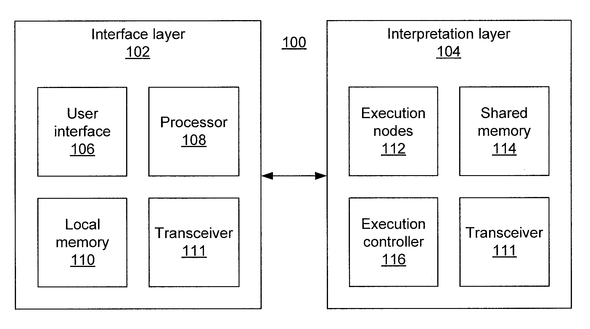
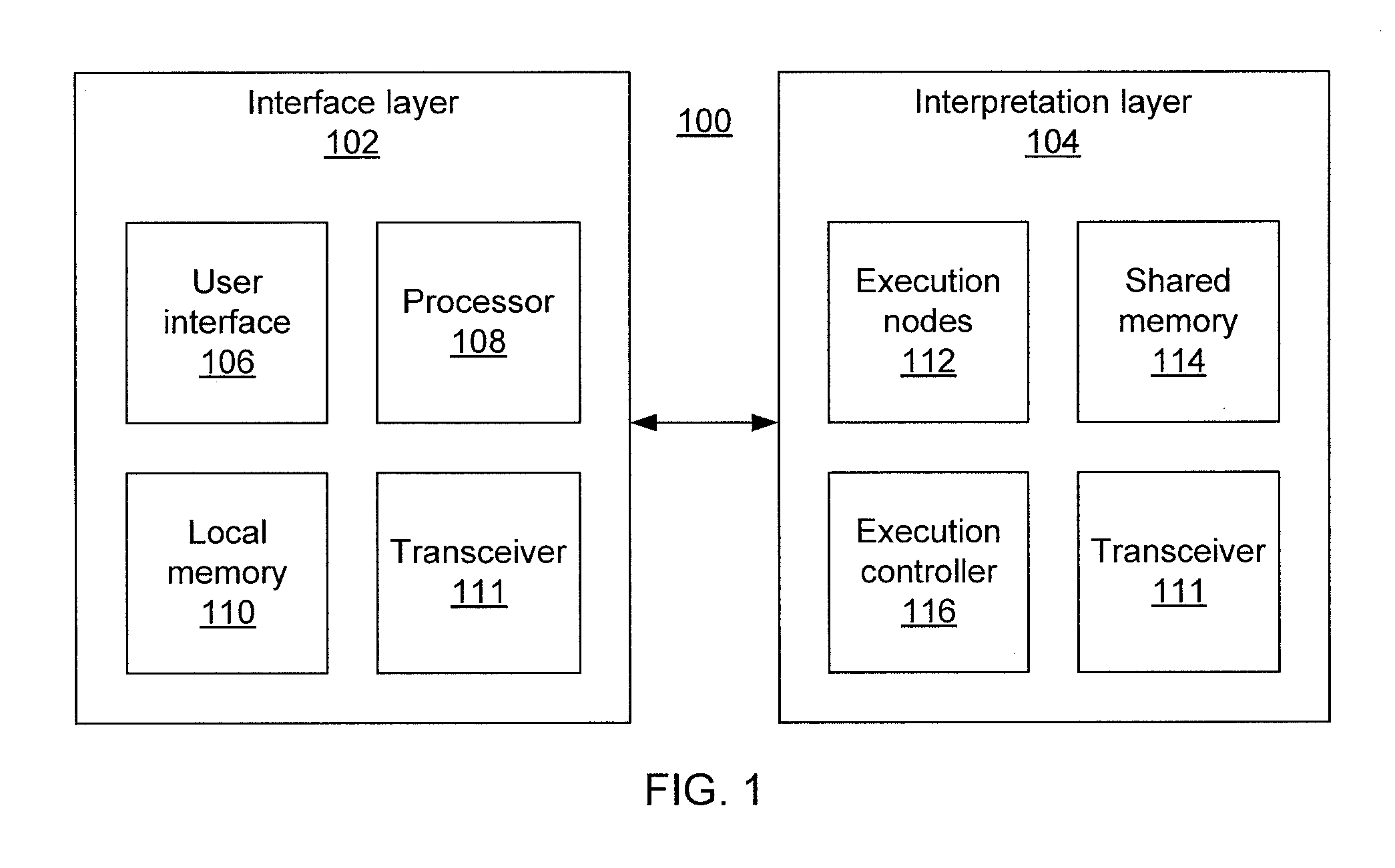
[0018]Referring now in detail to the figures in which like numerals represent the same or similar elements and initially to FIG. 1, an analytic system 100 according to the present principles is shown. An interface layer 102 includes a user interface 106, a processor 108, and local memory 110. The user interface 106 may be...


PUM


Abstract

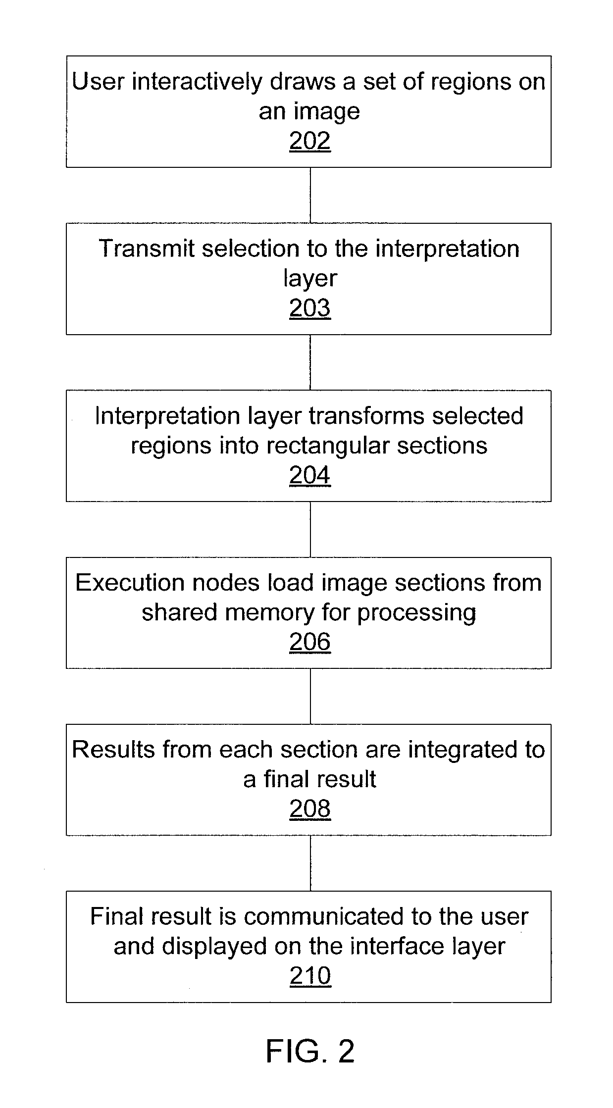
Methods and systems for interactive image analysis include receiving a selection of a region of an image and a request for analysis of the selection at an interface layer, transferring the selection and the request to an interpretation layer for analysis, dividing the selected region of the image into a plurality of sub-sections optimized for parallel computation to provide an analysis result that minimizes perceptible delay between receiving the request and receipt of results, analyzing the sub-sections using one or more execution nodes using a copy of the image stored in a shared memory, and providing combined analysis results to the interface layer for display.

Description

RELATED APPLICATION INFORMATION[0001]This application claims priority to provisional application Ser. No. 61 / 514,143, filed on Aug. 2, 2011, to provisional application Ser. No. 61 / 514,144, filed Aug. 2, 2011, and to provisional application Ser. No. 61 / 514,146, filed Aug. 2, 2011, each incorporated herein by reference. This application is related to application Ser. No. TBD, Attorney Docket No. 11027 (449-238) entitled, “DIGITAL PATHOLOGY SYSTEM WITH LOW-LATENCY ANALYTICS,” filed concurrently herewith and incorporated herein by reference. This application is related to application Ser. No. TBD, Attorney Docket No. 11028 (449-239) entitled, “CLOUD-BASED DIGITAL PATHOLOGY,” filed concurrently herewith and incorporated herein by reference.BACKGROUND[0002]1. Technical Field[0003]The present invention relates to digital pathology and, more particularly, to image analysis performed with whole-slide imaging.[0004]2. Description of the Related Art[0005]Pathologists and medical doctors analyz...

Claims


Application Information

Patent Timeline
Patent Type & Authority Applications(United States)
IPC IPC(8): G06K9/34
CPCG06K9/00979G06V10/95
Inventor COSATTO, ERICMALON, CHRISTOPHERGRAF, HANS P.
Owner NEC CORP
Learn More