Lung ventilation device

a ventilation device and a technology for lung, applied in the direction of audible signalling system, operating means/releasing devices for valves, instruments, etc., can solve the problems of complex design, affecting the visualization of patient data, and affecting the patient's diagnosis, etc., to facilitate and promote effective patient monitoring and surveillance, improve visualization and recognition, and enhance patient safety levels

Inactive Publication Date: 2013-05-23
INTERMED EQUIPAMENTO MEDICO HOSPITALAR
View PDF9 Cites 15 Cited by
  • Summary
  • Abstract
  • Description
  • Claims
  • Application Information

AI Technical Summary

Benefits of technology

[0027]Therefore, the purpose of the present invention is to provide a lung ventilation device that comprises a graphical interface configured in a manner that promotes effective patient monitoring and surveillance, being endowed with technical and functional characteristics that safely and efficiently resolve the problems and limitations of the equipment of the prior art.
[0028]More particularly, the purpose of the present invention is to provide a lung ventilation device that comprises a man-machine interface that can facilitate and promote effective patient monitoring and surveillance when caregiver is distant from the bed, improving the visualization and recognition of the main control parameters and the respective alarms, in order to enhance patient safety levels.
[0029]More particularly, the purpose of the present invention is to provide a lung ventilation device fitted with a patient monitoring and surveillance interface, where the main parameters associated with the patient are presented in a way that allows easy visualization at distance and interpretation of the clinical status of the patient, considerably reducing the cognitive load imposed on the medical staff members, thus considerably enhancing patient surveillance and safety.
[0030]Furthermore, the purpose of the present invention is to provide a lung ventilation device fitted with a graphical interface comprising the means for facilitating the detection of the occurrence of critical alarms, even by an operator away of the patient.

Problems solved by technology

It should be noted that the graphics do not usually allow fast and easy patient diagnosis.
Beyond the graphics, the interfaces of the equipment of the prior art also present large number of information, including numerical data, messages, controls and alarms, whose complexity hampers their visualization, interpretation and analyses for taking decisions.
In addition to the inconvenient aspects mentioned above, another extremely relevant aspect is that the graphical interfaces of the equipment of prior the art are not properly designed for visualization at distance, requiring medical staff members to approach to the lung ventilation device, and it is only then that they can see the available information, including alarm status.
Furthermore, in case of a problem with the patient or the equipment, these interfaces were designed to trigger visual and audible alarms, although not adequately highlighting the visual indicator, due to the problems mentioned above.
Thus, it is impossible for healthcare agents to distinguish between two alarms with the same priority but requiring different levels of urgency, such as disconnection of the respiratory circuit, which could lead to death or a rise in airway pressure because the patient coughed, with no major consequences.
Therefore, the patients are monitored at a working distance that does not allow adequate visualization of the graphical interface, far less any identification of the main associated parameters and alarms, due to the configuration of the graphical interface and the disposition of the parameters presented.
It is important to stress that, in these environments, and due to the distribution of the above mentioned monitored beds by the medical staff, merely identification of which alarm, as well as the item of equipment and the patient, becomes an extremely arduous task, which is often difficult to complete immediately, imposing cognitive loads on the staff members that exceed acceptable limits.
This difficulty or limitation on detecting a dangerous condition considerably increases the risk of harm to patients.
Occurs that some alarms classified under the regulatory standards as highly critical require faster responses than others, with the patient at risk of death.
Difficulties and resulting delays in meeting these situations greatly increase the risks involved.
In other words, due to the configuration of the interfaces, as well as constant alarms sounding simultaneously in the monitored environments, medical staff members do not have an efficient tool available that allows fast and immediate identification of endangered patients, as well as the reason why the alarm was triggered.
In conclusion, the interfaces have been designed to allow the operator to adjust the equipment, providing the largest possible amount of information on the screen, but they have clearly not been designed to allow and / or facilitate effective monitoring and surveillance of patients at the usual distance of the medical staff work.
Several patents describe ventilator interfaces including means to make them more user friendly, although it is noted that all of them provide means to assist ventilator setting but not effective patient vigilance.
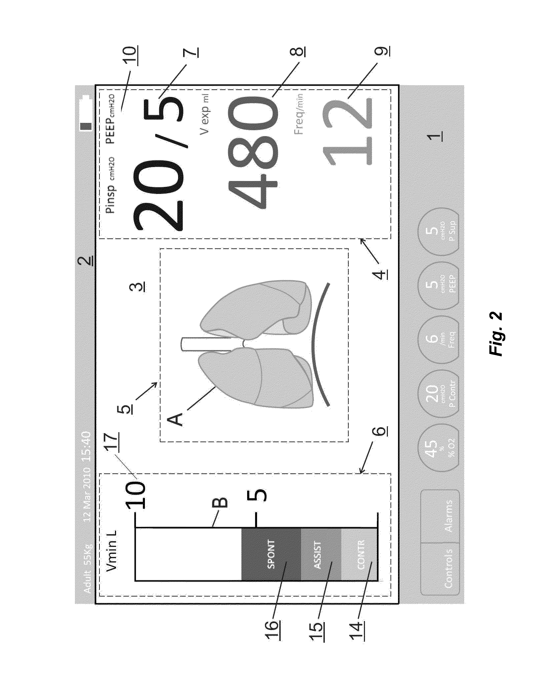
However, due to the scale of the Figure, as well as the anatomical characteristics of each patient (sex, age, weight, pathology, height, etc.), it is not possible to obtain an adequate correlation and a resolution between the effective volume received by the patient and the alteration in size associated with the lung.
This absence of accurate information provided by the stylized lung image requires the association of other numerical and graphical data that, according to the prior filing in question, are associated on the same screen, increasing the cognitive load of the medical staff, together with difficulties in visualization and the complexity of the analyses for diagnosis.
As may be noted, the graphical interfaces of the equipment of the prior art disclose a complex configuration that is difficult for the medical staff to see, particularly when positioned at distance of the beds, meaning that it is not possible to engage in any immediate clinical analysis without walking over to the respective bed.
Furthermore, a simple correlation between a stylized animated image and the patient requires considerable cognitive effort, when attempting to obtain more accurate and reliable information on the clinical status of the patient.
The conventional safety system, with visual and audible alarms integrated to the control and monitoring interface, imposes high cognitive load on the healthcare staff, hampering the recognition and response to a deleterious situation for the patient.
Furthermore, the graphical interfaces of the equipment of the prior art do not allow the immediate detection of the critical aspects of monitored patients at usual wok distances.

Method used


Image

  • Lung ventilation device
  • Lung ventilation device
  • Lung ventilation device

Examples


Embodiment Construction

[0037]In order to ensure an easier understanding of the configurable elements of the graphical interface of the lung ventilation device, according to the present invention, as well as its preferred embodiments, the numerical references will not be repeated in full in all the Figures, as this might hamper the understanding of some details illustrated therein.

[0038]FIG. 1 schematically represents a typical ventilation system of the type contemplated by the present invention, comprising a flow and pressure control valve 23 connected to a gas source 31, which controls the inspiratory flow through an inflow tube 27 connected to the patient 32 through a “Y” shaped connector 30. The patient 32 exhales the gas through an outflow tube 29, connected to the other end of the “Y” shaped connector 30, as controlled by exhalation valve 24. The airway pressure of the patient is transmitted from the “Y” shaped connector 30 which is connected to a pressure transducer 26 through a tube 28.

[0039]The in...


PUM


Abstract

The present invention refers to a lung ventilation device fitted with a patient monitoring and surveillance graphical interface comprising technical and functional characteristics that can reduce the cognitive load of the medical staff members in environments whose monitored beds require safe and efficient surveillance, principally for the identification and detection of parameters that are monitored at distance. More particularly, the present invention comprises a graphical interface whose configuration allows visualization at distance and interpretation of the main parameters associated with the patient, as well as includes means for facilitating the detection of the occurrence of critical alarms, even by an operator away from the patient.

Description

PRIORITY CLAIM AND CROSS-REFERENCE TO A RELATED APPLICATION[0001]This application claims priority of Brazilian Patent Application Number PI 1102142-0 filed May 20, 2011, the entire contents of which are incorporated by reference herein.TECHNICAL FIELD[0002]The present invention refers to a lung ventilation device fitted with a patient monitoring and surveillance graphical interface comprising technical and functional characteristics that can enhance patient surveillance and safety levels. More particularly, the graphical interface of the lung ventilation device comprises a configuration developed in a manner whereby the main parameters associated with the patient are presented in a way that allows easy visualization and interpretation at distance, lessening the cognitive load on the medical staff, and enhancing patient surveillance and safety. Furthermore, the present invention encompasses means for facilitating the detection of the occurrence of critical alarms, principally by an o...

Claims


Application Information

Patent Timeline
Patent Type & Authority Applications(United States)
IPC IPC(8): A61M16/00
CPCA61M16/0057A61M16/0051A61M16/12A61M2016/0027A61M2016/0039A61M2205/8212A61M2202/0208A61M2205/505A61M2205/581A61M2205/584A61M2205/8206A61M2016/0042A61M16/024
Inventor BONASSA, JORGECALVO LONARDONI, JOSE AUGUSTOSANTAREM DE ANDRADE, ORLANDO LUIZ
Owner INTERMED EQUIPAMENTO MEDICO HOSPITALAR
Why Eureka
Try Eureka