Method, reagent, and apparatus for detecting a chemical chelator

a chemical chelator and reagent technology, applied in biochemistry apparatus and processes, material analysis through optical means, instruments, etc., can solve the problems of insufficient cost-effectiveness, inability to monitor risk, and inability to use regularly outside the laboratory. , conventional laboratory based techniques requiring culturing of microorganisms are accurate, and are not suitable for general use to monitor hygien

Inactive Publication Date: 2013-05-23
ADVANCED BIOMEDICAL
View PDF11 Cites 6 Cited by
  • Summary
  • Abstract
  • Description
  • Claims
  • Application Information

AI Technical Summary

Benefits of technology

[0028]If necessary, the spores may be disrupted as a preliminary step in order to release DPA into the sample as described previously. However, the applicants have found that this is usually unnecessary as the reagent comprising the dye and the metal ion penetrates the envelope of many bacterial spores and so can be used to stain spores. Thus in a particular embodiment, preliminary disruption of the spores is unnecessary. This is a surprising discovery given the aforementioned difficulties of staining spores. Furthermore, by causing a dye to be released upon contact with the DPA, the method of the present invention can be used to reduce the background fluorescence and thereby solves the poor contrast problems of simply detecting calcium with dyes. It also solves the weak signal problems associated with the use of luminescence from a metal ion. It also allows the luminescence of the dye to be detected together with the luminescence from the metal ion, providing yet further selectivity. Samarium, europium, terbium and dysprosium ions can be used to detect many chelates, including DPA, where energy transfer from the chelates to the rare-earth ion provides strong luminescent signal amplification. The luminescent signal can be measured using time gated detection in order to reduce interference from background fluorescent signals which decay far more rapidly than the luminescent signal. Therefore luminescent signal may be measured as fluorescence life-time. The luminescent signal may also be measured as shift in wavelength of emission or excitation.
[0029]Importantly, by causing a dye to be released upon contact with the DPA, it allows a compartment (such as the core or the cortex of the spore) that is rich in DPA to be preferentially stained. The invention also allows a stained spore compartment to be imaged with an imaging system such as a microscope. The ability to preferentially stain a DPA rich compartment within the spore is believed to have important implications for researching and exploiting the microbiology of spores. In particular, it can be used to provide selective treatment for bacterial endospores since if the spore can be stained, it can be made photosensitive, and thus can be germinated or destroyed by illuminating with light.
[0030]DPA is an effective chelator of lanthanides and other metal ions, is more effective than other chelators that may be present in the cell wall of vegetative cells including the mother cell that forms the endospore, and in viable spores is present in much higher concentrations. Accordingly, higher staining of spores is observed than the staining of the mother cell, or other cells that will be present in the sample. This is important because it allows specific detection of bacterial endospores compared to vegetative bacterial cells, fungal spores, pollen, and viruses, none of which contain DPA.
[0070]The reagent may comprise a luminescent dye and a metal ion complex, the metal ion is bound to the luminescent dye, the luminescence of the dye is quenched by the metal ion, the reagent being characterized in that the metal ion is capable of binding to the DPA, and the dye and the metal ion are such that the DPA can compete for the metal ion in competition with the dye, thereby dequenching the luminescence of the dye.
[0086]The invention also provides an endospore characterised in that it has been treated by permeating a reagent comprising a luminescent dye bound to a metal ion into the endospore, which endospore comprises a spore coat, at least one compartment, dipicolinic acid or a derivative thereof, the luminescent dye, and a plurality of the metal ion, wherein the spore coat surrounds the compartment, the compartment contains the dipicolinic acid, the luminescent dye has a characteristic luminescence when excited by the optical radiation, the metal ions when bound to the luminescent dye reduce the luminescence of the luminescent dye and increase the rate of diffusion of the luminescent dye through the spore coat; the metal ions bind to the dipicolinic acid in preference to binding to the luminescent dye; the luminescence of the dye is increased when the metal ion is released from the dye and binds to the dipicolinic acid, and the luminescent dye within the endospore luminesces when excited by optical radiation.

Problems solved by technology

Given the large number of pathogens which pose health risks it is expensive to monitor risk from every pathogen using a highly specific test.
Conventional laboratory based techniques requiring culturing of microorganisms are accurate but are not rapid or sufficiently cost-effective to be used regularly outside the laboratory.
Such tests as colony counting and the polymerase chain reaction (PCR) are thus not suited for general use to monitor hygiene.
These tests are however relatively insensitive and costly.
The ATP test however has a number of limitations.
The ATP assay with its above said disadvantages is incapable of detecting spores.
This is a disadvantage because the presence of a small number of spores can be infectious.
However, because markers such as calcium, nucleic acids and phospholipids are naturally abundant, they are not specific indicators of spore presence.
Calcium in particular is not only found in most life forms, but is also frequently present (for example in hard water) making calcium a poor marker for detecting spores.
This mechanism is not highly efficient due to non-radiative decay.
A neutral ligand like trioctyl phosphine oxide has been used as a secondary ligand to obtain a better quantum yield, but with limited success.
None of these chemical modifications are likely to solve the problems of detecting native DPA in spores.
The molar absorbtivities and quantum yields of DPA-metal chelates are poor.
Such complexes do not yield as bright a fluorescence signal when compared to fluorescent dyes that are commonly used in high sensitivity assays.
Another limitation of the standard DPA-Tb3+ assay for detecting spores is that it generally requires the DPA to be released from spores into solution which is then measured to record luminescence.
Another limitation of the DPA-Tb3+ assay is that the complex requires excitation energy by ultra-violet light, typically at 276 nm.
This brings a number of problems when considering biological samples and the matrices they may be present in, since most such samples will themselves have ultra-violet absorption properties.
In addition to this, microscopic applications of this assay are severely limited as ultra-violet light sources for microscopic imaging are expensive and pose health risks to the observer.
While such dyes can provide much more sensitive and highly stable signals, they have not found any use in direct detection of DPA in spores as they have do not have the ability to bind DPA specifically.
However, calcium being very prevalent in biological samples is not a specific marker of spores.
Furthermore, calcein has not been used to penetrate into spores, or detect single spores.
The measurement thus provides very poor contrast between fluorescence emitted from calcein bound to the calcium from a spore and background fluorescence emerging from the calcein in solution.
The method is also non-specific as it is unable to detect DPA as calcein is not able to respond to DPA.
However, the resulting luminescent signal, that occurs by energy transfer from DPA to terbium, is weak, is not localized within the spore, requires UV excitation, and is not able to detect single spores.

Method used


Image

  • Method, reagent, and apparatus for detecting a chemical chelator
  • Method, reagent, and apparatus for detecting a chemical chelator
  • Method, reagent, and apparatus for detecting a chemical chelator

Examples


example 1

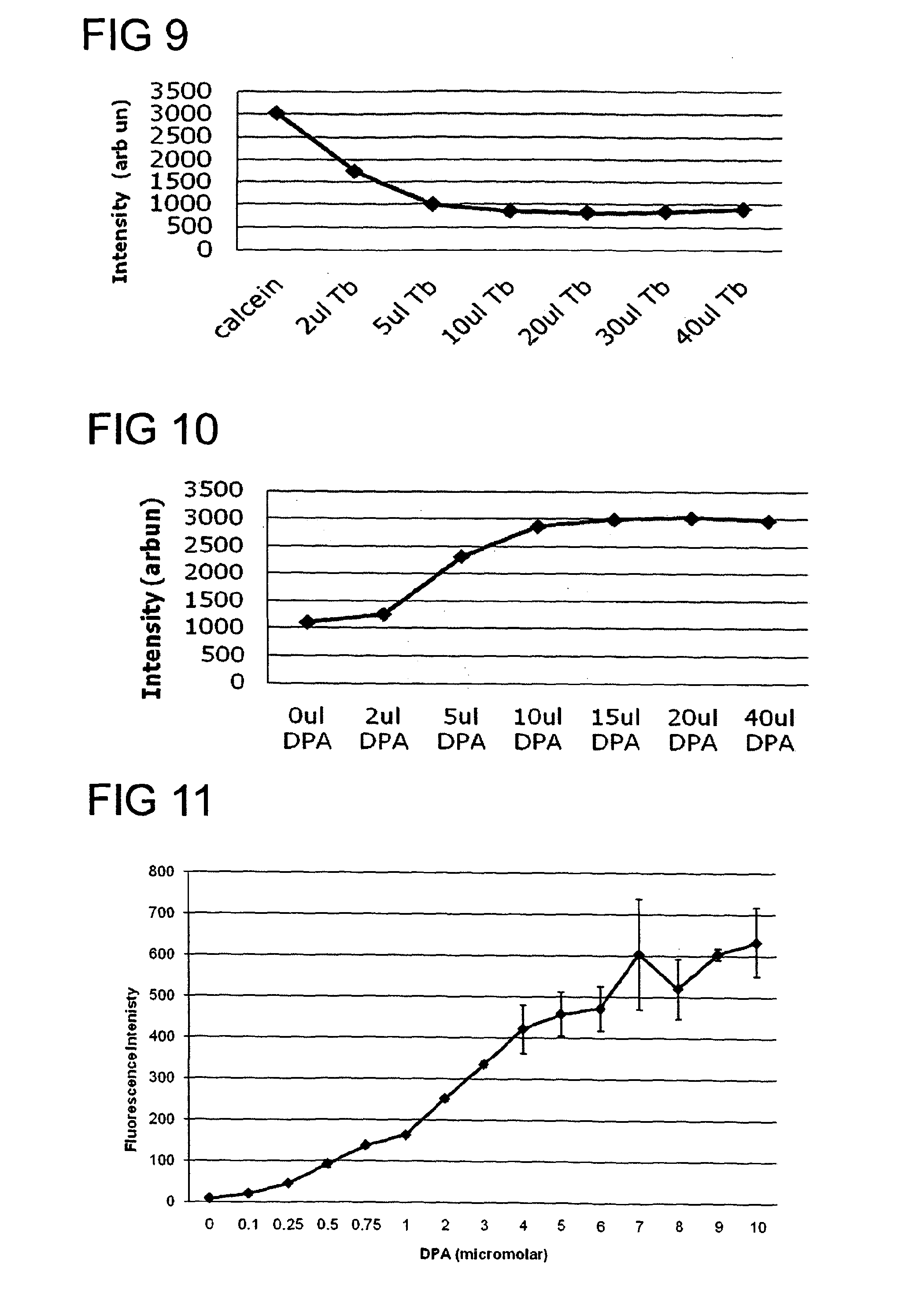
[0159]Several fluorescence dyes, including fluorescein, rhodamine and calcein, were screened for their ability to bind to zinc, cobalt, terbium, aluminium, and several other metal ions. Their fluorescence emission was recorded before and after adding the metal ion. Many of these show quenching of the fluorescence. One of the efficient dyes was calcein whose fluorescence was quenched by a number of different metal ions including ions of zinc, cobalt, iron and terbium. These ions were tested for their ability to bind to DPA. The binding ability to DPA is variable in terms of affinity. Preferably the metal ion and the dye are selected such that the DPA has a higher affinity to bind to the metal ion than the dye binding to the metal ion for sensitive detection. From this study, terbium and europium were found to be two of the most efficient metal ions in terms of their quenching effect on calcein and ease of dequenching with DPA. although many others were also shown to have potential. S...

example 2

[0160]Calcein and terbium (III) chloride hexahydrate, europium (III) chloride hexahydrate, cobalt (II) chloride hexahydrate, lead (II) chloride, calcium chloride and other general chemicals and solvents were purchased from Sigma-Aldrich (UK).

[0161]Calcein was selected as the luminescent dye 3, and terbium as the metal ion 4. The fluorescence emission signal 7 was recorded using an excitation wavelength (λex) 82 of calcein set at 485 nm and the emission wavelength (λem) 83 set at 520 nm. FIG. 9 shows the quenching of the fluorescent signal when a fixed concentration of calcein (1.2 μM) is titrated with various aliquots (μL) of terbium chloride (2 mM) solution. As more and more terbium chloride is added, the fluorescence signal drops. From this quenching curve a ratio of greater than four terbium ions per calcein molecule was found to quench the fluorescent signal efficiently. Therefore from such information, quenched solutions of calcein and terbium mixture could be prepared for test...

example 3

[0162]A dilute solution comprising calcein and terbium chloride in 1:8 molar ratio, respectively, was prepared in 10 mM TES buffer pH 7.4. Various amounts of DPA were titrated into this solution and changes in fluorescence intensity of calcein measured using an excitation wavelength of 485 nm and emission at 520 nm. FIG. 11 shows the dose response curve for DPA detection.

[0163]Further optimization of this assay and reagents including molar ratio and nature of dye and metal used may produce dose response curves with even higher sensitivity.

[0164]It is also possible to use the invention to detect chelators other than DPA. Different chelators may respond differently depending on their binding affinities to the metal. To illustrate how a suitable chelator may be identified the fluorescence emission spectrum of the calcein-terbium stain was recorded in the presence and absence of DPA, EDTA, Lactic acid, 2,6-Diaminopimelic acid (DAP), N-Acetylmuramic acid and acetic acid.

[0165]FIG. 31 sho...


PUM

PropertyMeasurementUnit
molar concentrationaaaaaaaaaa
volumeaaaaaaaaaa
excitation wavelengthaaaaaaaaaa

Abstract

A method for detecting a first chelator (shown in the figure as dipicolinic acid DPA (1), the method comprising the steps of providing a reagent (2) comprising a second chelator (shown as a luminescent dye (3) and a metal ion (4), contacting the reagent (2) with a sample (6) containing the DPA (1), exciting a luminescence (7) of the dye (3), and detecting the luminescence (7) emitted by the dye (3), the method being characterized in that the metal ion (4) is bound to the luminescent dye (3) within the reagent (2), the luminescence (7) of the dye (3) is altered by the metal ion (4), the metal ion (4) is capable of binding to the DPA (1), and the dye (3) and the metal ion (4) are such that the DPA (1) can compete for the metal ion (4) in competition with the dye (3), thereby re-establishing the luminescence (7) of the dye (3).

Description

FIELD OF INVENTION[0001]The invention relates to a method, reagent, and apparatus for detecting a chemical chelator and in particular dipicolinic acid (DPA). The invention has application in the detection or treatment of microbes including spores for medical, clinical, food hygiene, and military applications. Spores obtainable using the method form a further aspect of the invention.BACKGROUND TO THE INVENTION[0002]Monitoring of pathogens is becoming increasingly important to control infection both in clinical and community settings. Rapid tests are needed to ensure food, water and environment safety. Given the large number of pathogens which pose health risks it is expensive to monitor risk from every pathogen using a highly specific test. Thus non-specific monitoring of microbial contamination is now employed in hygiene monitoring. Conventional laboratory based techniques requiring culturing of microorganisms are accurate but are not rapid or sufficiently cost-effective to be used ...

Claims


Application Information

Patent Timeline
Patent Type & Authority Applications(United States)
IPC IPC(8): G01N21/76
CPCG01N21/763C12Q1/04C12Q1/22G01N21/6486G01N33/5008G01N33/5082
Inventor AOJULA, HARMESH SINGHCLARKE, DAVID JOHN
Owner ADVANCED BIOMEDICAL
Why Eureka
Try Eureka