Surgical Stapling Device Including A Camera

a stapling device and camera technology, applied in the field of surgical instruments including cameras, can solve the problems of time-consuming endoscope insertion and removal of staplers, and achieve the effect of reducing the anvil profile and reducing the trauma during removal

Inactive Publication Date: 2013-10-31
TYCO HEALTHCARE GRP LP
View PDF3 Cites 371 Cited by
  • Summary
  • Abstract
  • Description
  • Claims
  • Application Information

AI Technical Summary

Benefits of technology

[0010]The anvil assembly includes a tiltable anvil that provides a reduced anvil profile to reduce trauma during removal. The tiltable anvil tilts automatically when the device has been fired and unapproximated. A camera assembly is operatively coupled to a surface of the tiltable anvil. The camera ass

Problems solved by technology

This however requires removal of the stapler

Method used


Image

  • Surgical Stapling Device Including A Camera
  • Surgical Stapling Device Including A Camera
  • Surgical Stapling Device Including A Camera

Examples


Embodiment Construction

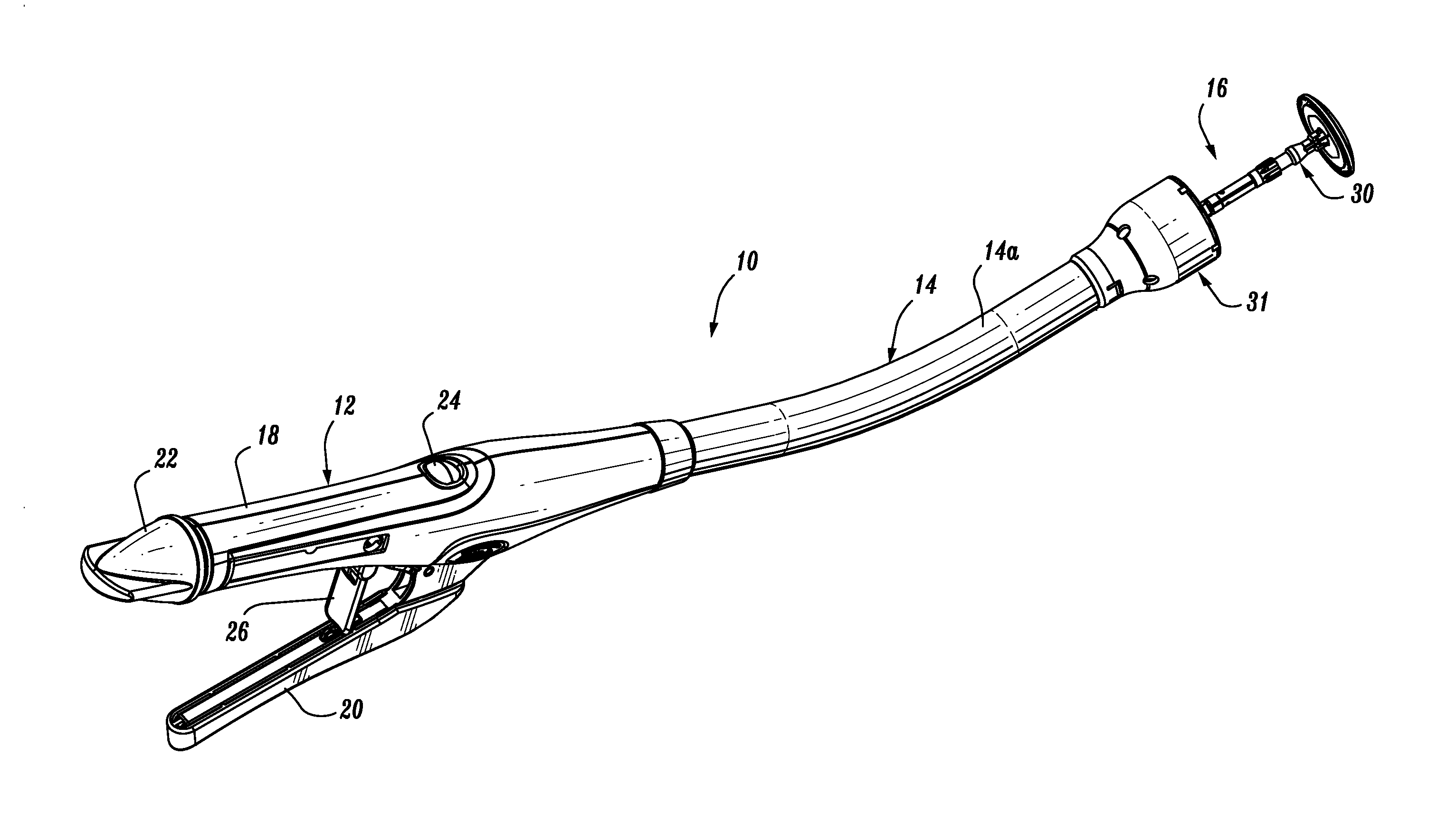
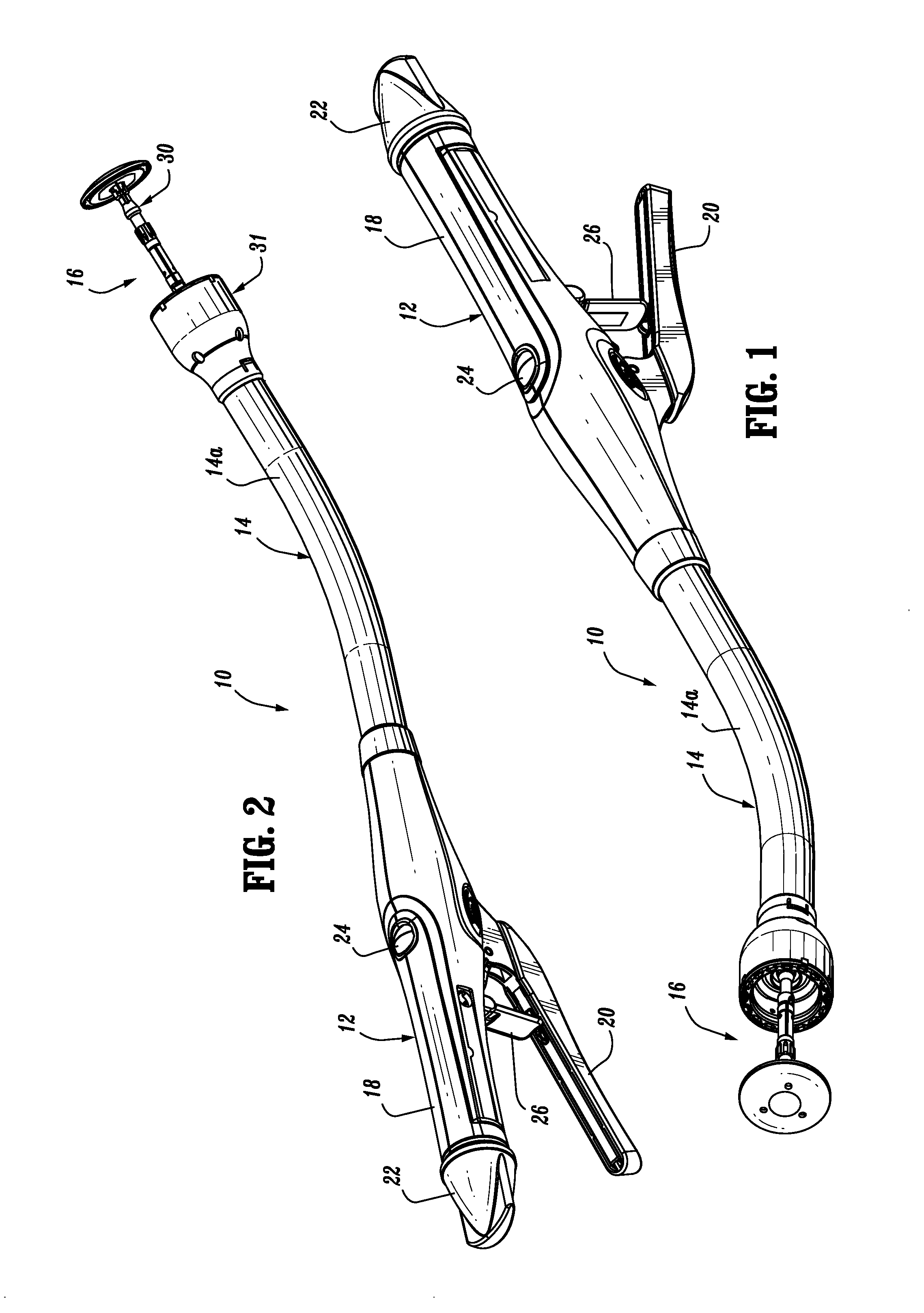
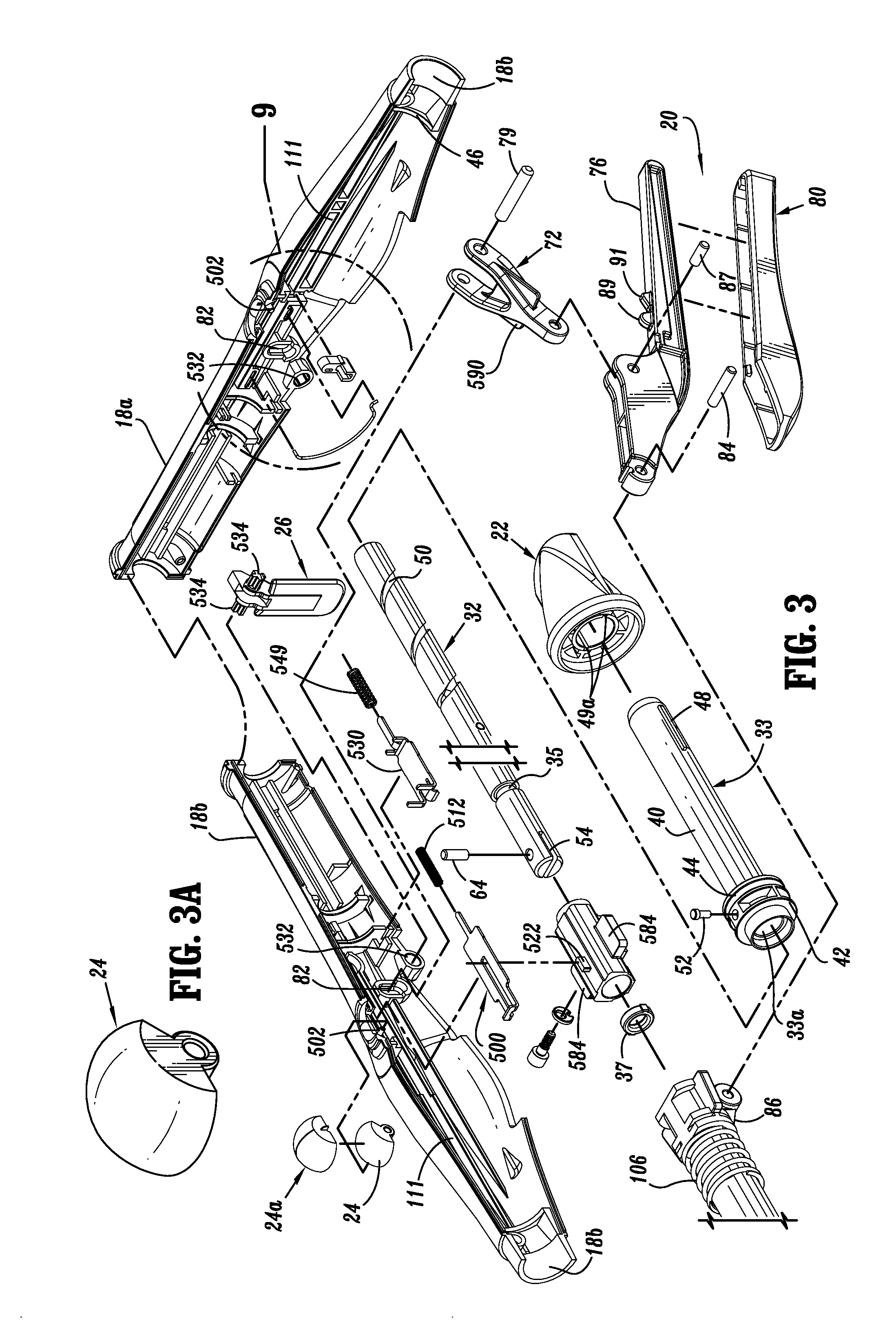
[0124]Embodiments of the presently disclosed surgical stapling device will now be described in detail with reference to the drawings in which like reference numerals designate identical or corresponding elements in each of the several views. Throughout this description, the term “proximal” will refer to the portion of the instrument closest to the operator and the term “distal” will refer to the portion of the instrument furthest from the operator.

[0125]FIGS. 1 and 2 illustrate one embodiment of the presently disclosed surgical stapling device shown generally as 10. Briefly, surgical stapling device 10 includes a proximal handle assembly 12, an elongated central body portion 14 including a curved elongated outer tube 14a, and a distal head portion 16. Alternately, in some surgical procedures, e.g., the treatment of hemorrhoids, it is desirable to have a substantially straight central body portion. The length, shape and / or the diameter of body portion 14 and head portion 16 may also ...


PUM

PropertyMeasurementUnit
Poweraaaaaaaaaa
Lightaaaaaaaaaa

Abstract

A surgical stapling apparatus including a handle assembly, an elongated shaft extending distally from the handle assembly, a shell assembly containing a plurality of staples arranged in an annular array, and a firing mechanism for advancing staples from the shell assembly. An anvil assembly including a tiltable anvil head movable from an aligned operative position to a tilted position. A camera assembly is operatively coupled to a distal surface of the tiltable anvil head such that an orientation of the camera assembly changes as the anvil head moves from the operative to the tilted position.

Description

CROSS-REFERENCE TO RELATED APPLICATIONS[0001]This application claims the benefit of and priority to U.S. Provisional Patent Application No. 61 / 638,533, filed Apr. 26, 2012, the entire disclosure of which is incorporated by reference herein.BACKGROUND[0002]1. Technical Field[0003]The present disclosure relates generally to a surgical instrument including a camera. More particularly, the present disclosure relates to a surgical circular stapling device for applying surgical staples to body tissue which device includes a camera.[0004]2. Background of Related Art[0005]Anastomosis is the surgical joining of separate hollow organ sections. Typically, an anastomosis procedure follows surgery in which a diseased or defective section of hollow tissue is removed and the remaining end sections are to be joined. Depending on the desired anastomosis procedure, the end sections may be joined by either circular, end-to-end or side-to-side organ reconstruction methods.[0006]In a circular anastomosi...

Claims


Application Information

Patent Timeline
IPC IPC(8): A61B1/04A61B17/068
CPCA61B1/04A61B17/068A61B17/1155A61B2017/00221A61B2017/00734A61B2017/07257A61B1/00087A61B1/00183A61B90/30A61B90/361A61B2090/3616
Inventor MA, YONG
Owner TYCO HEALTHCARE GRP LP
Why Eureka
Try Eureka