
Apparatus and methods for removing and collecting biopsy specimens from biopsy devices with fixation and preparation for histopathological processing or other analysis

a biopsy device and biopsy specimen technology, applied in medical science, surgical procedures, vaccination/ovulation diagnostics, etc., can solve the problems of contaminated specimens, minute specimen damage or loss, and loss of orientation and biopsy sequence, so as to facilitate the passage of the biopsy instrument

Inactive Publication Date: 2013-12-26
ZIMMON DAVID S
View PDF14 Cites 11 Cited by
  • Summary
  • Abstract
  • Description
  • Claims
  • Application Information

AI Technical Summary

Benefits of technology

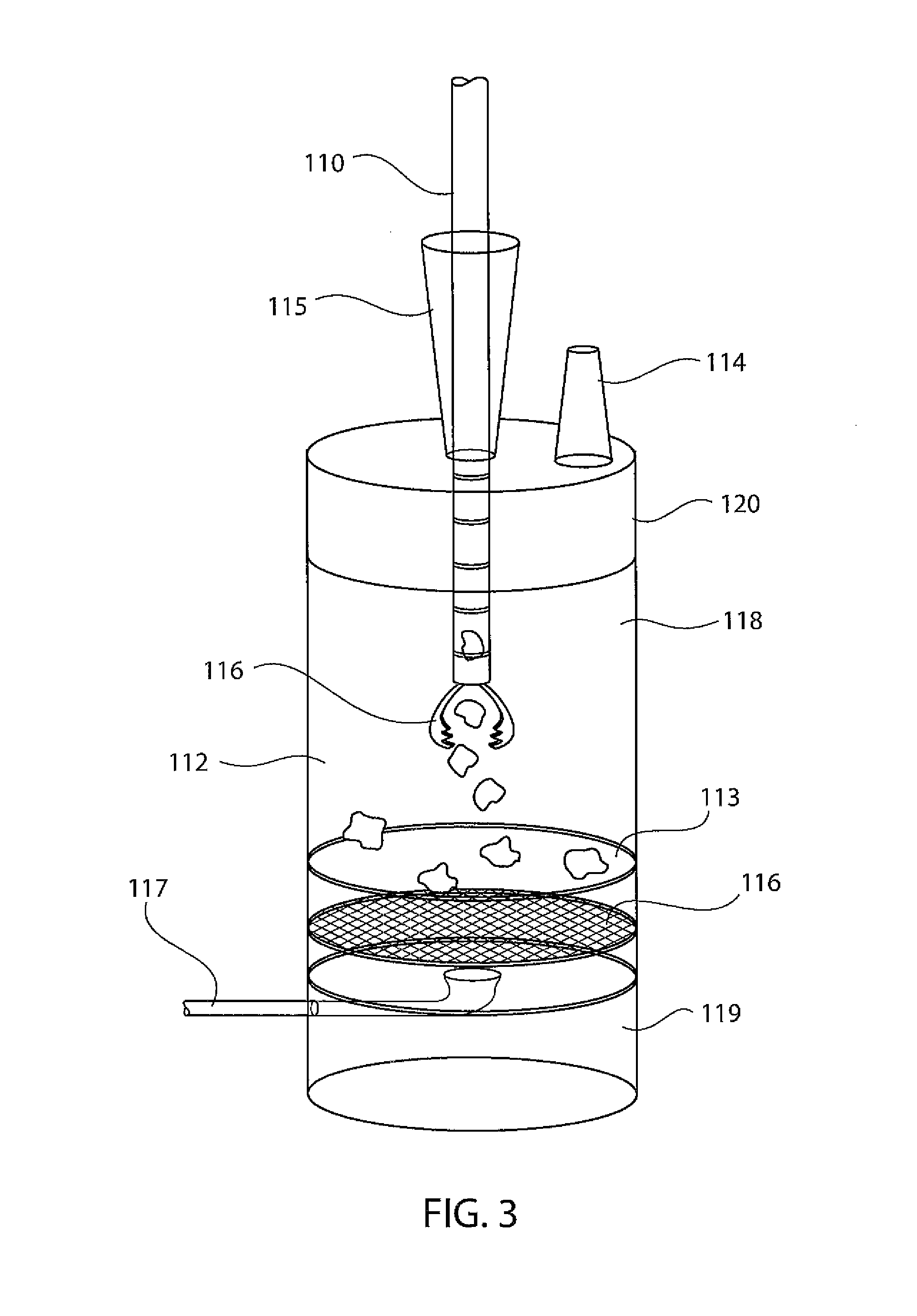
The present invention provides an apparatus and method for removing and collecting biopsy specimens from spring based multiple biopsy storage cylinders or other biopsy devices combined with fixation, fixative removal, and paper wrapping or collecting in a cassette for histopathological processing or other analysis. The invention minimizes manual specimen handling, limits staff exposure to sharps, infectious material, and toxic fixatives. The biopsy device can be washed and reused in the same patient. The invention also prepares biopsy specimens for processing by collecting them in a receptacle with a removable screen and a bottom drain for removing fixative. The invention improves the efficiency of the pathology laboratory by automating the process of retrieving, inspecting, and paper wrapping fixed or unfixed specimens for processing. The double cylinder embodiment is useful for narrow and flexible biopsy devices or when toxic fixatives require a second cylinder wall to guard against environmental contamination.

Problems solved by technology

Minute specimens may be damaged or lost during this process.
Specimens are contaminated during retrieval and lose orientation and biopsy sequence in the container.
During these operations staff is exposed to sharps, infectious material and toxic fixatives.
These actions prolong the operation and anesthesia with increased patient risk and cost.
During this process the minute specimens are subject to damage, fragmentation and loss.
These manual operations expose staff to infectious material and toxic fixatives.
This tedious, labor intensive process is expensive in staff required, time, equipment and laboratory space.
But in situ fixation and processing destroys the biopsy device preventing additional passes for collecting specimens.

Method used


Image

  • Apparatus and methods for removing and collecting biopsy specimens from biopsy devices with fixation and preparation for histopathological processing or other analysis
  • Apparatus and methods for removing and collecting biopsy specimens from biopsy devices with fixation and preparation for histopathological processing or other analysis
  • Apparatus and methods for removing and collecting biopsy specimens from biopsy devices with fixation and preparation for histopathological processing or other analysis

Examples


Embodiment Construction

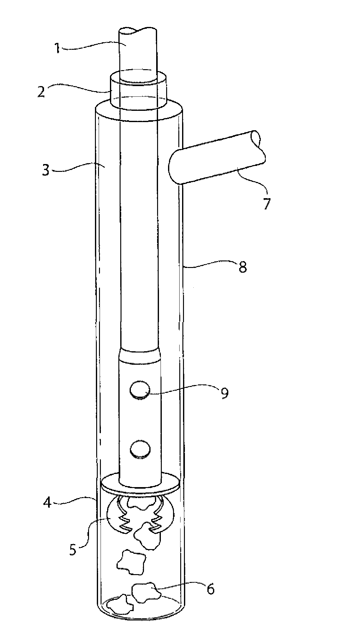
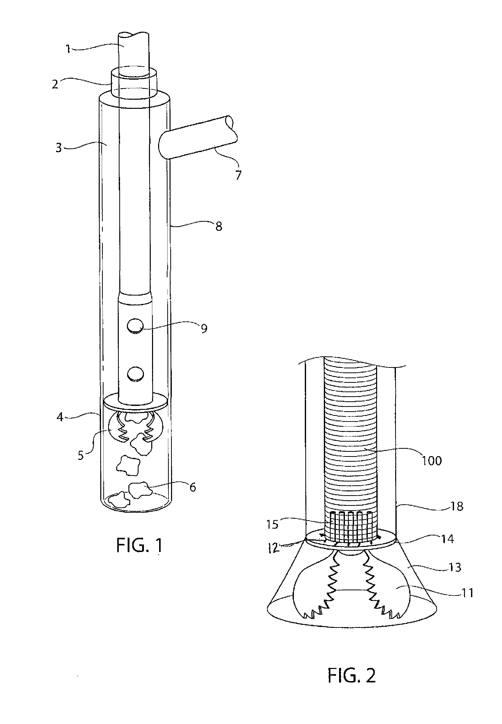
[0020]One embodiment of the invention as shown in FIG. 1 is a single wall clear plastic irrigating apparatus for collecting individual specimens or specimens groups from a spring based multiple biopsy storage cylinder or other biopsy devices. The closed biopsy device and shaft are passed through a proximal perforated elastic seal 2 down an irrigating apparatus lumen 3 and out the perforated distal seal 4. The spring based multiple biopsy device jaws are opened by activating the biopsy mechanism (not shown) to open an exit path for specimen 6 removal. Irrigating fluid or gas is injected through the side arm 7 into cylinder 8 of lumen 3.

[0021]Fluid passes into the lumen 3 through spring based multiple biopsy storage cylinder perforations 9 entraining the storage cylinder specimens 6 into a receptacle (not shown). After removing the specimens in the receptacle, the biopsy devices is washed by irrigating through the side arm 7 and removed ready for reuse.

[0022]FIG. 2 shows a forceps bio...


PUM


Abstract

An apparatus and method for removing biopsy specimens from spring based spring based multiple biopsy or forceps biopsy devices by irrigating or suctioning. The apparatus collects, fixes and prepares specimens for histopathologic processing limiting manual labor, workplace risk and time previously required. After removal from the biopsy device, specimens are fixed in a histopathological processing cassette or on specimen wrapping paper for transportation to the pathology laboratory. In pathology, fixative is drained and specimens inspected for gross description followed by paper wrapping and histopathological processing with minimum handling, exposure to infectious material or toxic fixatives. After removing the biopsy specimens, the biopsy device is washed in the apparatus to eliminate contaminants and removed, ready for another biopsy sequence. A spring based spring based multiple biopsy device using these methods can collect numerous biopsy batches from different sites with segregated processing and analysis of each batch.

Description

CROSS-REFERENCE TO RELATED APPLICATIONS[0001]This application claims priority under 35 USC 119(e) of U.S. Provisional Application Ser. No. 61 / 690,389, filed on Jun. 25, 2012.BACKGROUND OF THE INVENTION[0002]1. Field of the Invention[0003]The present invention relates to removing and collecting biopsy specimens from biopsy devices with fixing for analysis by histopathological, biological or physical methods. Biopsy specimens are minute pieces of fragile tissue that are difficult to remove from the biopsy device used to cut or scrape them from tissue. Biopsy specimens require careful handling to prevent loss, fragmentation and contamination. After manual removing from the biopsy instrument specimens require fixing and collecting for analysis. Fixed specimens must be removed from fixative, inspected and prepared for histopathological processing. This laborious process is slow and costly.[0004]Spring based spring based multiple biopsy devices take many biopsies in a single pass to the t...

Claims


Application Information

Patent Timeline
Patent Type & Authority Applications(United States)
IPC IPC(8): A61B10/02
CPCA61B10/02A61B10/0096
Inventor ZIMMON, DAVID S.
Owner ZIMMON DAVID S
Learn More