Vein imaging systems and methods

a vasculature and system technology, applied in the field of system and method for imaging a patient vasculature, can solve the problems of difficult to determine the exact location of the blood vessel, difficult to place the distal tip of the needle in the blood vessel lumen, and difficulty in proper placement of hypodermic and procedural needles, so as to facilitate the detection of infiltration or extravasation within the target area

Inactive Publication Date: 2014-02-06
EVENA MEDICAL
View PDF12 Cites 160 Cited by
  • Summary
  • Abstract
  • Description
  • Claims
  • Application Information

AI Technical Summary

Benefits of technology

[0008]Various embodiments disclosed herein can relate to a system for facilitating detection of infiltration or extravasation within a target area on a body portion of a patient. The system can include a light source configured to direct light onto the target area, a light sensor configured to receive light from the target area and to generate an image of the target area, and a display configured to display the image of the target area. The system can be configured such that the displayed image shows the presence of infiltration or extravasation when infiltration or extravasation is present in the target area.

Problems solved by technology

The exact location of the blood vessel may be difficult to determine because it is not in the direct sight of the medical practitioner attempting to gain vascular access.
Placing the distal tip of the needle in the blood vessel lumen may also be difficult for similar reasons.
Consequently, proper placement of hypodermic and procedural needles can be challenging.
Furthermore, because the patient's vasculature is not readily visible, it is often difficult for a medical practitioner to determine whether a patient's blood vessel has been compromised (e.g., due to vein collapse, vein blockage, vein leakage, etc.).
If medical fluids are infused (e.g., via an IV connection) into a compromised blood vessel, the fluid can leak out of the blood vessel and into the surrounding tissue, resulting in infiltration or extravasation, which can cause damage to the surrounding tissue and can prevent infused medication from properly entering the patient's vasculature.
Thus, it can be difficult for the medical practitioner to determine whether a blood vessel has been compromised, especially for low amounts of infiltration or extravasation.
Also, in some instances, fluid can leak from an underside of the blood vessel (e.g., facing generally away from the surface of the skin), which can cause infiltration or extravasation that is relatively deep in the patient's tissue and is more difficult to detect using conventional patency checks.

Method used


Image

  • Vein imaging systems and methods
  • Vein imaging systems and methods
  • Vein imaging systems and methods

Examples


Embodiment Construction

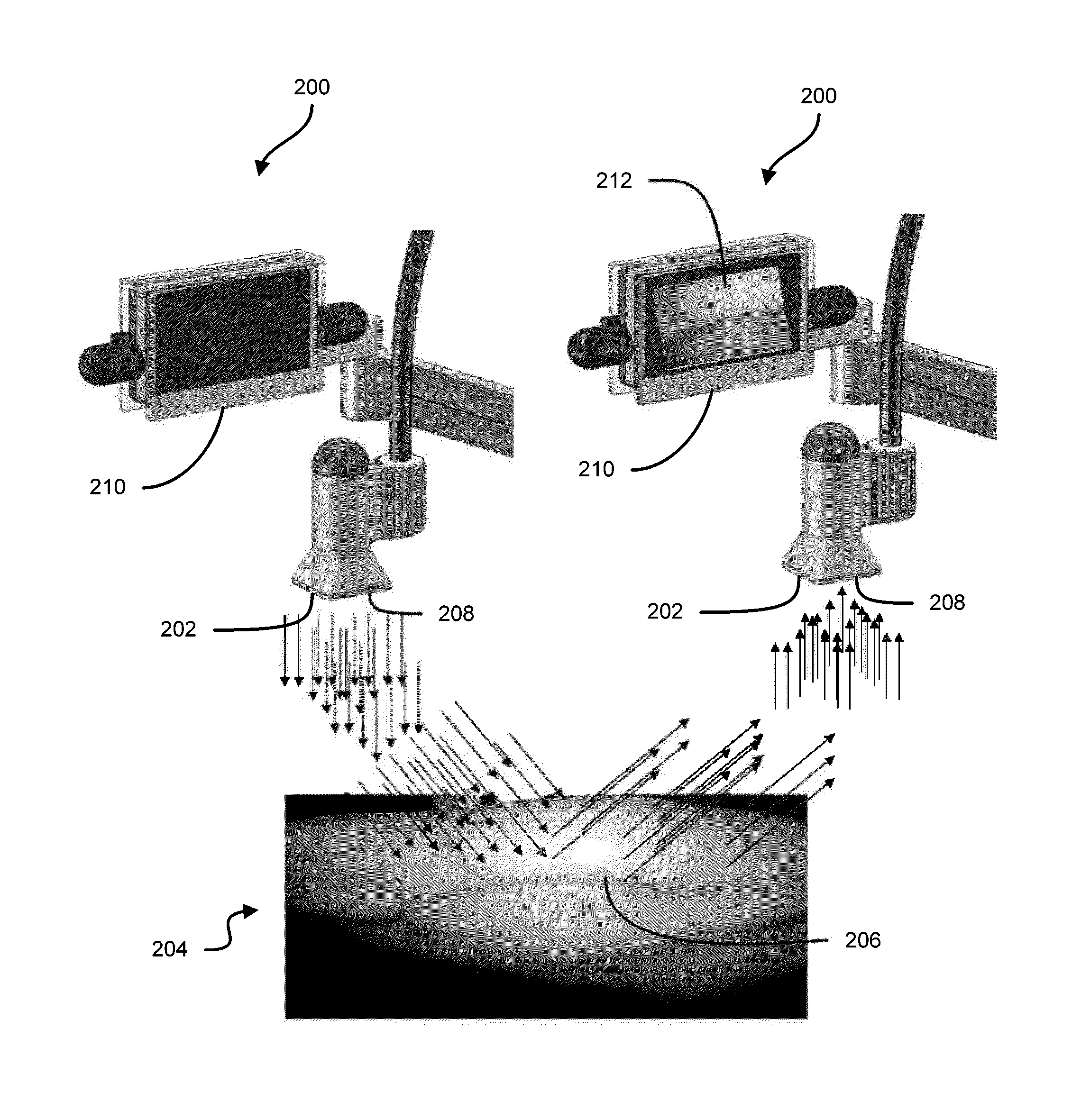
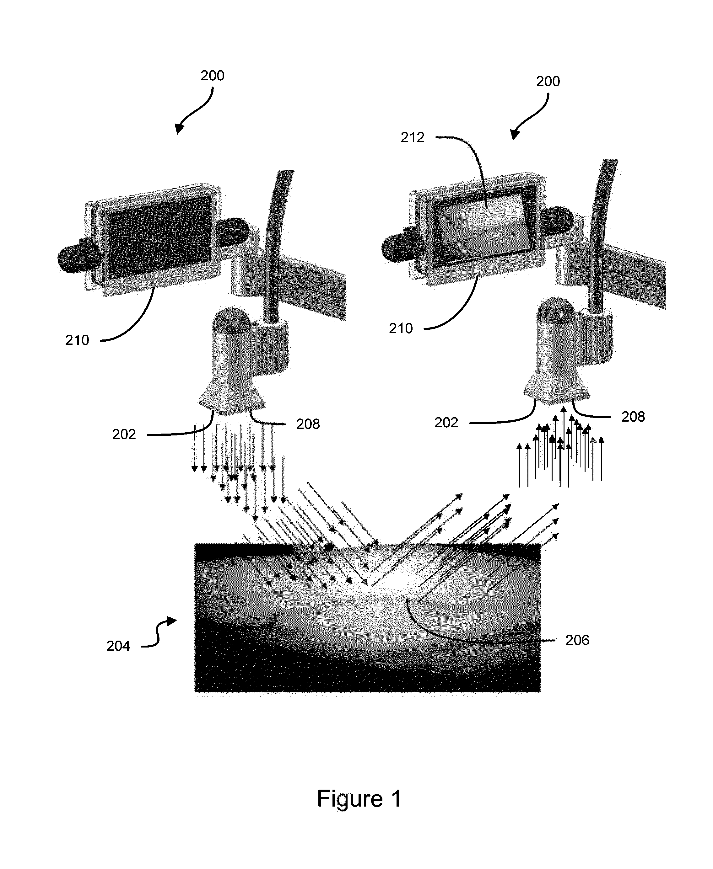
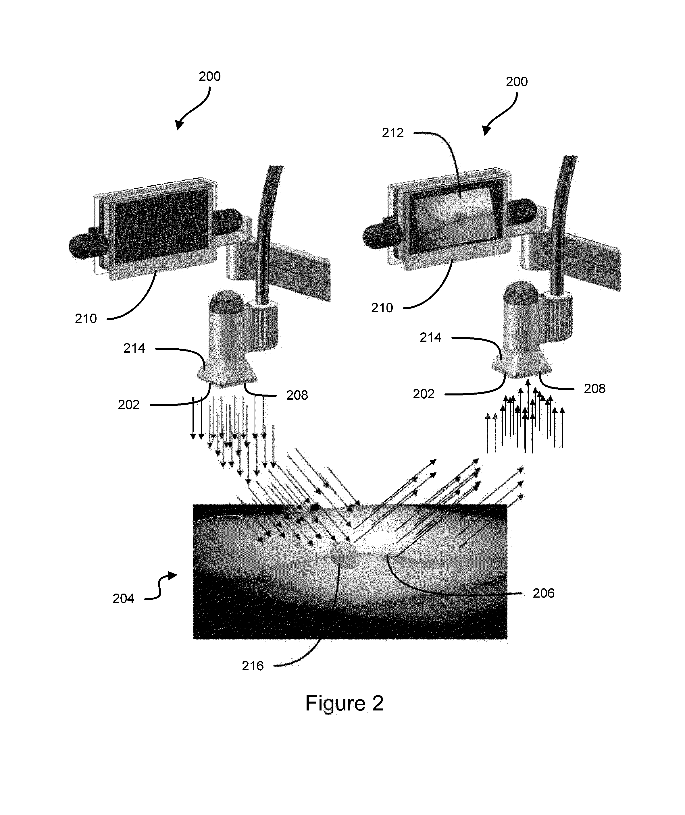
[0158]FIG. 1 shows an imaging system 200 that can be used to view the patent's vasculature and / or to identify infiltration or extravasation. The system can include a light source 202, such as an array of light emitting diodes (LEDs), that is configured to emit light onto a target area 204, such as an arm or other portion of a patient's body that includes one or more blood vessels 206 (e.g., veins). Although various embodiments are described herein with connection to viewing of veins, various features and methods described herein can be used for imaging other blood vessels and for imaging other vessels (e.g., lymphatic vessels) that transfer bodily fluids other than blood. The light source 202 can emit wavelengths of light that cause less of the light to be reflected or scattered by the veins and more of the light to be reflected by the tissue surrounding the veins. As used herein, the term “reflected” light includes light that is scattered. For example, near infrared (NIR) light can...


PUM


Abstract

Some embodiments of this disclosure relates to systems and methods for imaging a patient's vasculature. For example, near infrared (NIR) light can be used to illuminate a target area and light that is reflected or scattered from the target area can be used for generate an image of the target area. In some embodiments, the system can be configured such that the image shows the presence, absence, or extent of infiltration or extravasation in the target area. The system can be configured to document that presence, absence, or extend of infiltration or extravasation at an infusion site. In some embodiments, an imaging system can be mounted onto a patient so that the imaging system can monitor an infusion site, and the imaging system can be configured to automatically detect the presence of infiltration or extravasation.

Description

CROSS-REFERENCE TO RELATED APPLICATIONS[0001]This application claims the benefit under 35 U.S.C. §119(e) of U.S. Provisional Patent Application No. 61 / 639,012 (Attorney Docket No. EVENA.001PR), filed Apr. 26, 2012, and titled VEIN IMAGING SYSTEMS AND METHODS, U.S. Provisional Patent Application No. 61 / 639,808 (Attorney Docket No. EVENA.001PR2), filed Apr. 27, 2012, and titled VEIN IMAGING SYSTEMS AND METHODS, and U.S. Provisional Patent Application No. 61 / 714,684 (Attorney Docket No. EVENA.013PR), filed Oct. 16, 2012, and titled VEIN IMAGING SYSTEMS AND METHODS, each of which is hereby incorporated by reference in its entirety and made a part of this specification for all that it discloses.BACKGROUND[0002]1. Field of the Disclosure[0003]Some embodiments of this disclosure relate to systems and methods for imaging a patient's vasculature, such as to facilitate the insertion of an intravenous line or to facilitate assessment of a blood vessel, an infusion site, or a target area on a p...

Claims


Application Information

Patent Timeline
Patent Type & Authority Applications(United States)
IPC IPC(8): A61B5/00
CPCA61B5/0075A61B5/742A61B5/0059A61B5/02007A61B5/4833A61B5/6803A61M5/16836A61M2005/1588A61M2205/15A61M2205/3306A61M2209/04A61B5/0013A61B5/0077A61B5/489A61B5/6891A61B5/14546A61B5/14551G16H10/60G16H30/20G16H20/17A61B5/7282A61B5/7445
Inventor HARRIS, MELVYN L.HARRIS, TONI A.BALL, FRANK J.GRUEBELE, DAVID J.CESPEDES, IGNACIO E.
Owner EVENA MEDICAL
Why Eureka
Try Eureka