
Boosted consensus classifier for large images using fields of view of various sizes

a large image and consensus technology, applied in image enhancement, image analysis, instruments, etc., can solve the problems of inability to accurately and efficiently quantify image analysis tools, inability to accurately reflect the level of malignancy or heterogeneity based on isolated fovs, and inability to accurately reflect the level of malignancy or heterogeneity

Active Publication Date: 2014-03-06
RUTGERS THE STATE UNIV
View PDF12 Cites 34 Cited by
  • Summary
  • Abstract
  • Description
  • Claims
  • Application Information

AI Technical Summary

Benefits of technology

The invention describes a method for analyzing large, heterogeneous images using a boosted multi-field-of-view (FOV) framework. This method involves dividing the image into multiple FOVs at different fixed sizes, and detecting objects in each FOV. The method then generates a combined class decision based on the detected objects in each FOV, using a boosted multi-FOV classifier. The method can be applied to various types of images, such as microscopic images or histopathologic images, and can be particularly useful for analyzing cancer images. The technical effect of this invention is to provide a more efficient and accurate method for analyzing large, heterogeneous images.

Problems solved by technology

Yet clinical usage of the BR grading system is often limited by concerns about intra- and inter-rater variability.
A possible reason for such variability is that pathologists currently lack the automated image analysis tools to accurately and efficiently quantify BR grade in histopathology.
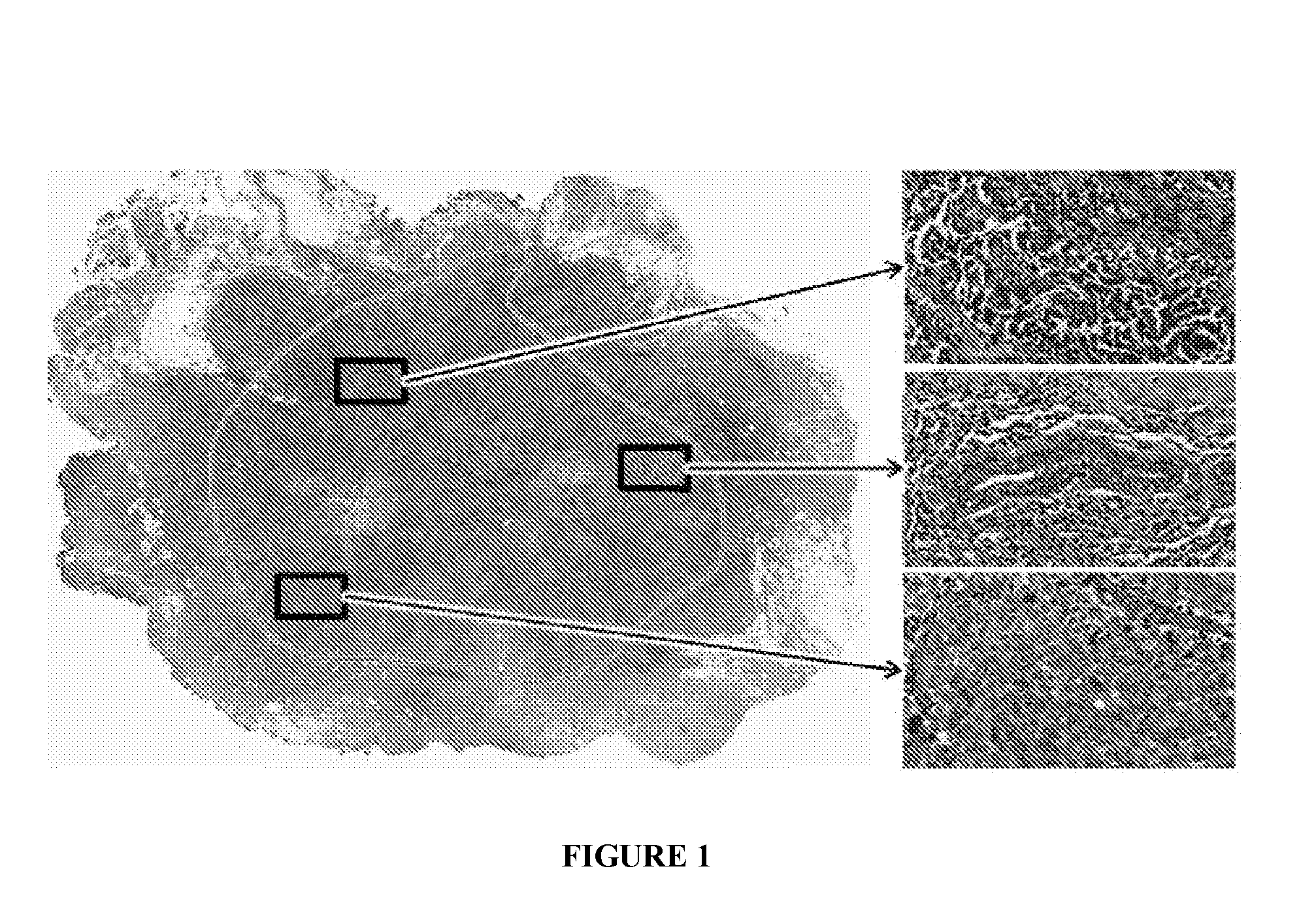
This step is important due to the heterogeneous nature of BCa, whereby predictions based on isolated FOVs may not accurately reflect the level of malignancy or heterogeneity in an entire histopathology slide, as described in Connor et al.
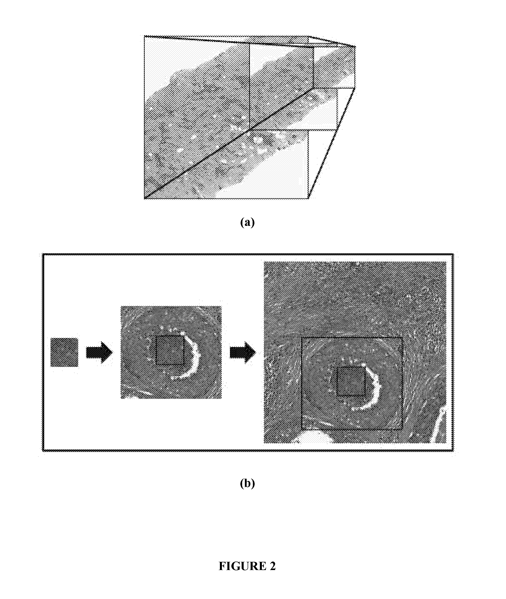
While clinicians perform this task implicitly, the a priori selection of an optimal FOV (i.e. image patch) size for computerized analysis of entire histopathology slides is not straightforward.
However, analyzing domain-specific image architecture is more challenging, since it remains invariant to changes in scale (although our visual perception and ability to detect objects within the image will vary).
However, they are inherently serial processes that cannot leverage integration techniques (e.g. consensus) to more effectively combine predictions returned by C0, C1, and C2.
Although clinicians perform this task implicitly, the a priori selection of an optimal FOV (i.e. image patch) size for computerized analysis of an entire histopathology slide is not straightforward.

Method used


Image

  • Boosted consensus classifier for large images using fields of view of various sizes
  • Boosted consensus classifier for large images using fields of view of various sizes
  • Boosted consensus classifier for large images using fields of view of various sizes

Examples


example 1

Evaluation of the Boosted, Multi-Field-of-View (Multi-FOV) Classifier to Distinguish Breast Cancer Patients of Differential Grading by Nuclear Architecture Extraction

[0256]This example demonstrates the ability of the boosted, multi-field-of-view (multi-FOV) classifier of the present invention to distinguish patients with low and high grades in breast cancer (BCa) histopathology images.

Dataset

[0257]Hematoxylin and eosin stained histopathology slides from 55 patients (36 low grade, 19 high grade) at the Hospital of the University of Pennsylvania were digitized via a whole slide scanner at 10× magnification (1 μm / pixel resolution). Each slide C is accompanied by (1) annotations from an expert pathologist denoting large regions of interest containing invasive cancer and (2) Bloom-Richardson (BR) grade labels denoting low (BR 3-5) or high (BR 7-9) grade. A wide range of FOV sizes (T={250, 500, 1000, 2000, 4000} pixels) was selected for analysis.

[0258]FIG. 6 shows exemplary breast cancer ...

example 2

Evaluation of the Boosted, Multi-Field-of-View (Multi-FOV) Classifier to Distinguish Breast Cancer Patients of Differential Grading by Nuclear Architecture Extraction and Nuclear Texture Extraction

[0267]This example demonstrates the ability of the boosted, multi-field-of-view (multi-FOV) classifier of the present invention to distinguish patients with low vs. high BR grade, low vs. intermediate BR grade, and intermediate vs. high BR grade in breast cancer (BCa) histopathology images.

Dataset

[0268]BCa histopathology slides were obtained from 126 patients (46 low BR, 60 intermediate BR, 20 high BR) at the Hospital of the University of Pennsylvania and The Cancer Institute of New Jersey. All slides were digitized via a whole slide scanner at 10× magnification (1 μm / pixel resolution). Each slide was accompanied by BR grade as determined by an expert pathologist. Commonly accepted clinical cutoffs used to define the low (BR 3-5), intermediate (BR 6-7), and high (BR 8-9) grade classes were...

experiment 1

Whole Slide Classification Using Nuclear Architecture

[0269]The ability of the boosted, multi-field-of-view (multi-FOV) classifier to discriminate entire BCa histopathology slides based on BR grade via architectural features {circumflex over (f)}NA was evaluated. Since the multi-FOV classifier utilizes a trained classifier, it is susceptible to the arbitrary selection of training and testing data. A 3-fold cross-validation scheme was used to mitigate such bias by splitting the data cohort into 3 subsets in a randomized fashion, from which 2 subsets are used for training and the remaining subset is used for evaluation. The subsets were subsequently rotated until a class prediction H(D; {circumflex over (f)}NA)ε{0,1} was made for each slide. A slide was said to be correctly classified if the class prediction matches the ground truth label (i.e. BR grade) assigned to the slide. The entire cross-validation procedure was repeated 20 times and mean classification results were reported.


PUM


Abstract

A system and method for predicting disease outcome by analyzing a large, heterogeneous image by a boosted, multi-field-of-view (FOV) framework, based on image-based features from multi-parametric heterogeneous images, comprises (a) inputting the heterogeneous image; (b) generating a plurality of FOVs at a plurality of fixed FOV sizes, the method for generating the plurality of FOVs at a plurality of fixed FOV sizes comprising dividing simultaneously, via the computing device, the large, heterogeneous image into (i) a plurality of FOVs at a first fixed FOV size from among the plurality of fixed FOV sizes; and (ii) a plurality of FOVs at a second fixed FOV size from among the plurality of fixed FOV sizes; (c) producing simultaneously for the heterogeneous image a combined class decision for: (i) the plurality of FOVs at the first fixed FOV size, and (ii) the plurality of FOV s at the second fixed FOV size.

Description

CROSS-REFERENCE TO RELATED APPLICATIONS[0001]This application claims priority to U.S. provisional application No. 61 / 431,286, filed on Jan. 10, 2011, titled “Boosted Consensus Classifier for Large Images using Fields of View of Various Sizes”, and U.S. provisional No. 61 / 435,607, filed on Jan. 24, 2011, titled “Boosted Consensus Classifier for Large Images using Fields of View of Various Sizes”, the entire disclosure of which is incorporated herein by reference.FIELD OF THE INVENTION[0002]The described invention relates to methods and systems of predicting classification grading, disease aggressiveness and outcome.BACKGROUND OF THE INVENTION[0003]Breast cancer (BCa) is an increasingly common cancer diagnosis in women. Advancements in screening, diagnostic, and therapeutic techniques in BCa have improved survival rates in recent years. (Jemal, A. et al., “Declining death rates reflect progress against cancer,”PLoS One, vol. 5, no. 3, p. e9584, 2010. Available at dx.doi.org / 10.1371 / jo...

Claims


Application Information

Patent Timeline
IPC IPC(8): G06T7/00G06V10/426G06V10/774
CPCG06T7/0012G06T2207/20072G06T2207/30024G06T2207/30068G06T7/12G06T7/149G06V20/698G06V10/426G06V2201/03G06V10/774G06F18/214
Inventor MADABHUSHI, ANANTBASAVANHALLY, AJAY
Owner RUTGERS THE STATE UNIV
Learn More