Ultrasound imaging system and method

a technology of ultrasound imaging and ultrasound, applied in the field of ultrasound imaging system and method, can solve the problems of inaccurate quantitative measurement values, patient heart rate changes, and difficulty in identifying the cardiac phase with complete certainty

Inactive Publication Date: 2014-05-08
GENERAL ELECTRIC CO
View PDF8 Cites 12 Cited by
  • Summary
  • Abstract
  • Description
  • Claims
  • Application Information

AI Technical Summary

Problems solved by technology

However, if the patient has an abnormal cardiac cycle or if the imaging plane is not properly located, it can be very difficult to identify the cardiac phase with complete certainty based solely on a B-mode image.
However, this technique poses a problem if the patient's heart rate changes between the acquisition of the u...

Method used


Image

  • Ultrasound imaging system and method
  • Ultrasound imaging system and method
  • Ultrasound imaging system and method

Examples


Embodiment Construction

[0014]In the following detailed description, reference is made to the accompanying drawings that form a part hereof, and in which is shown by way of illustration specific embodiments that may be practiced. These embodiments are described in sufficient detail to enable those skilled in the art to practice the embodiments, and it is to be understood that other embodiments may be utilized and that logical, mechanical, electrical and other changes may be made without departing from the scope of the embodiments. The following detailed description is, therefore, not to be taken as limiting the scope of the invention.

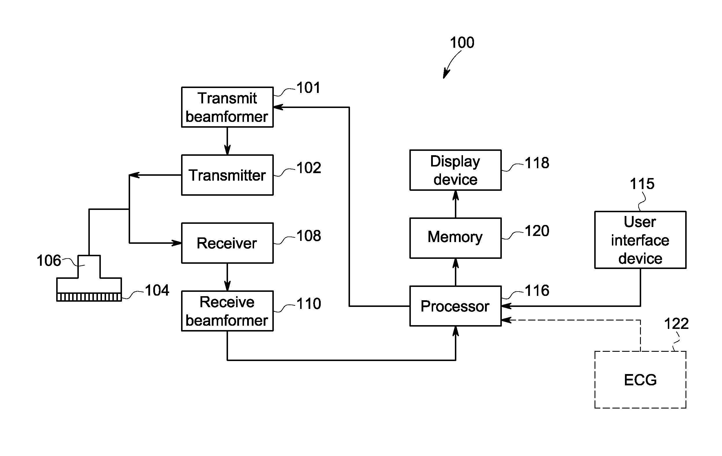
[0015]FIG. 1 is a schematic diagram of an ultrasound imaging system 100 in accordance with an embodiment. The ultrasound imaging system 100 includes a transmit beamformer 101 and a transmitter 102 that drive elements 104 within a probe 106 to emit pulsed ultrasonic signals into a body (not shown). The probe 106 may be an 2D array probe according to an embodiment. The pulsed ul...


PUM


Abstract

An ultrasound imaging system and method includes receiving a first ultrasound image of a region-of-interest (ROI) and associated first ECG data, the first ultrasound image including an M-mode image or a spectral Doppler image. The system and method includes receiving a cine loop of B-mode images acquired from the ROI and second associated ECG data. The system and method includes selecting a first phase and displaying, at the same time, a first one of the B-mode images at the first phase, the first ultrasound image, and a marker. The marker is positioned at a first position with respect to the first ultrasound image, the first position indicating the first phase.

Description

FIELD OF THE INVENTION[0001]This disclosure relates generally to an ultrasound imaging system and method for displaying an M-mode image or a spectral Doppler image at the same time as a B-mode image. A marker is displayed with the M-mode image or the spectral Doppler image. The position of the marker with respect to the M-mode image or the spectral Doppler image indicates the phase of the B-mode image.BACKGROUND OF THE INVENTION[0002]In echocardiography, there are many tasks and measurements that require identifying an ultrasound image from a very specific portion of the cardiac cycle. For example, it is commonly desired to obtain a volume or other measurement from a specific portion of the cardiac cycle such as aortic valve closure, aortic valve opening, mitral valve closure, or mitral valve opening. According to conventional techniques, a clinician would typically perform a visual assessment of B-mode images that are part of a cine loop in order to manually identify the desired ca...

Claims


Application Information

Patent Timeline
IPC IPC(8): G09G5/00G16H30/40
CPCA61B8/463G09G2380/08A61B8/0883A61B8/469A61B8/486A61B8/488A61B8/5284G16H30/40
Inventor LYSYANSKY, PETER
Owner GENERAL ELECTRIC CO
Why Eureka
Try Eureka