
System and Method for Registering Pre-Operative and Intra-Operative Images Using Biomechanical Model Simulations

a biomechanical model and simulation technology, applied in image analysis, image enhancement, instruments, etc., can solve the problems of large organ deformation, large organ deformation, and sliding between the viscera and abdominal wall

Active Publication Date: 2014-05-15
SIEMENS HEALTHCARE GMBH
View PDF5 Cites 31 Cited by
  • Summary
  • Abstract
  • Description
  • Claims
  • Application Information

AI Technical Summary

Benefits of technology

The present invention provides a way to accurately align pre-operative and intra-operative images of a target organ using a biomechanical model of abdomen deformation under gas insufflation. This is done by optimizing a measure of intensity similarity between the two images and estimating patient-specific parameters for insufflation. The technical effects of this invention include improved accuracy and efficiency in image-based surgery procedures.

Problems solved by technology

During laparoscopic surgery, organ surfaces can only be viewed through laparoscopic images and it is difficult to visualize sub-surface tumors.
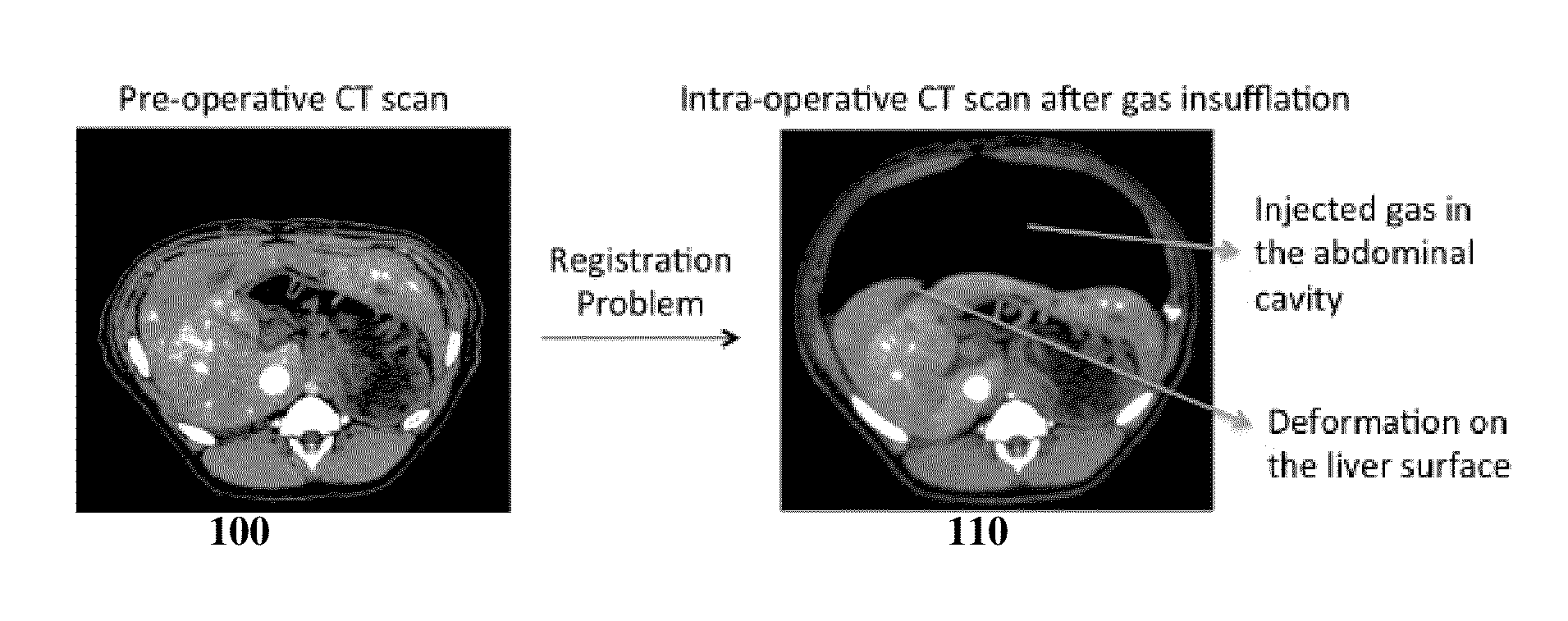
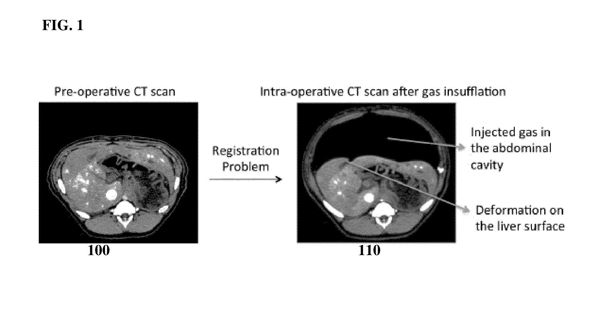
However, such registration is challenging due to gas insufflation and other surgical preparation, which can result in large organ deformation and sliding between the viscera and abdominal wall.
Thus, standard non-rigid image registration techniques do not provide accurate results.

Method used


Image

  • System and Method for Registering Pre-Operative and Intra-Operative Images Using Biomechanical Model Simulations
  • System and Method for Registering Pre-Operative and Intra-Operative Images Using Biomechanical Model Simulations
  • System and Method for Registering Pre-Operative and Intra-Operative Images Using Biomechanical Model Simulations

Examples


Embodiment Construction

[0013]The present invention relates to registration of pre-operative and intra-operative images of a target organ using biomechanical model simulations. Embodiments of the present invention are described herein to give a visual understanding of the methods for registering pre-operative and post-operative images using biomechanical model simulations. A digital image is often composed of digital representations of one or more objects (or shapes). The digital representation of an object is often described herein in terms of identifying and manipulating the objects. Such manipulations are virtual manipulations accomplished in the memory or other circuitry / hardware of a computer system. Accordingly, is to be understood that embodiments of the present invention may be performed within a computer system using data stored within the computer system.

[0014]Laparoscopic surgery is a minimally invasive procedure that is widely used for treatment of cancer and other diseases. During the procedur...


PUM


Abstract

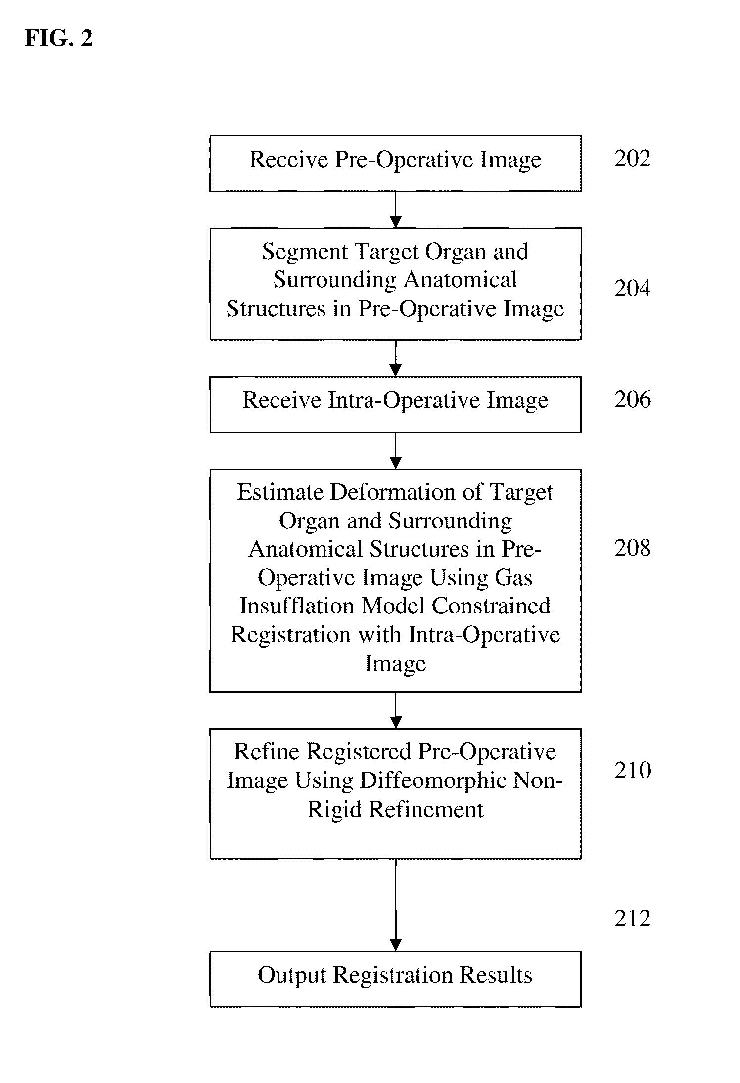
A method and system for registering pre-operative images and intra-operative images using biomechanical simulations is disclosed. A pre-operative image is initially registered to an intra-operative image by estimating deformations of one or more segmented anatomical structures in the pre-operative image, such as the liver, surrounding tissue, and the abdominal wall, using biomechanical gas insufflation model constrained. The initially registered pre-operative image is then refined using diffeomorphic non-rigid refinement.

Description

[0001]This application claims the benefit of U.S. Provisional Application No. 61 / 726,687, filed Nov. 15, 2012, the disclosure of which is herein incorporated by reference.BACKGROUND OF THE INVENTION[0002]The present invention relates to registration of pre-operative and intra-operative images for laparoscopic surgery, and more particularly to registration of pre-operative and intra-operative images of a target organ using biomechanical model simulations.[0003]Laparoscopic surgery is a minimally invasive procedure that is widely used for treatment of cancer and other diseases. Advantages, such as reduced post-operative pain and shorter recovery time, make laparoscopic surgery preferable to open surgery. During laparoscopic surgery, organ surfaces can only be viewed through laparoscopic images and it is difficult to visualize sub-surface tumors. Accordingly, intra-operative three dimensional (3D) images can be introduced to provide updated information during the laparoscopic surgery. ...

Claims


Application Information

Patent Timeline
Patent Type & Authority Applications(United States)
IPC IPC(8): G06T11/00
CPCG06T11/005G06T7/33G06T2207/10081G06T2207/30028G06T2207/30056
Inventor OKTAY, OZANZHANG, LI
Owner SIEMENS HEALTHCARE GMBH
Learn More