Luminal background cleaning

a technology of background cleaning and illumination, applied in the field of medical image processing, can solve the problems of many 11-regularized problems still remaining difficult to solv

Active Publication Date: 2014-05-22
SYNC RX
View PDF13 Cites 26 Cited by
  • Summary
  • Abstract
  • Description
  • Claims
  • Application Information

AI Technical Summary

Benefits of technology

[0041]Some applications of the present invention are applied to medical procedures performed, in whole or in part, on or within luminal body structures or body cavities. For some applications, a

Problems solved by technology

Despite this recent attention, many 11-regularized problems still remain

Method used


Image

  • Luminal background cleaning
  • Luminal background cleaning
  • Luminal background cleaning

Examples


Embodiment Construction

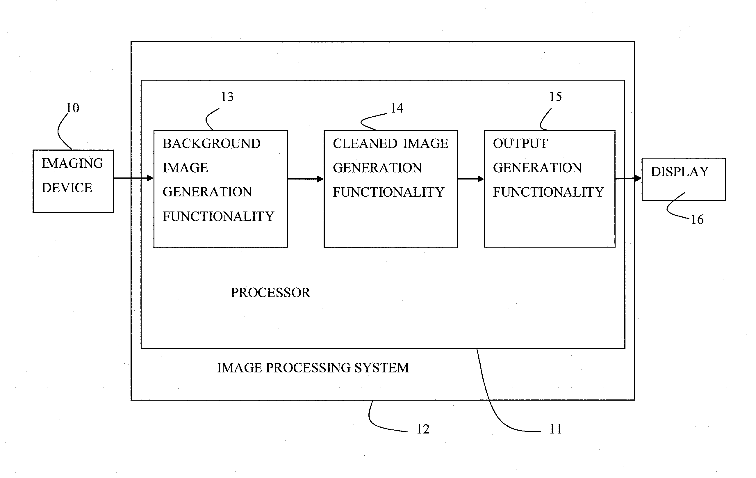
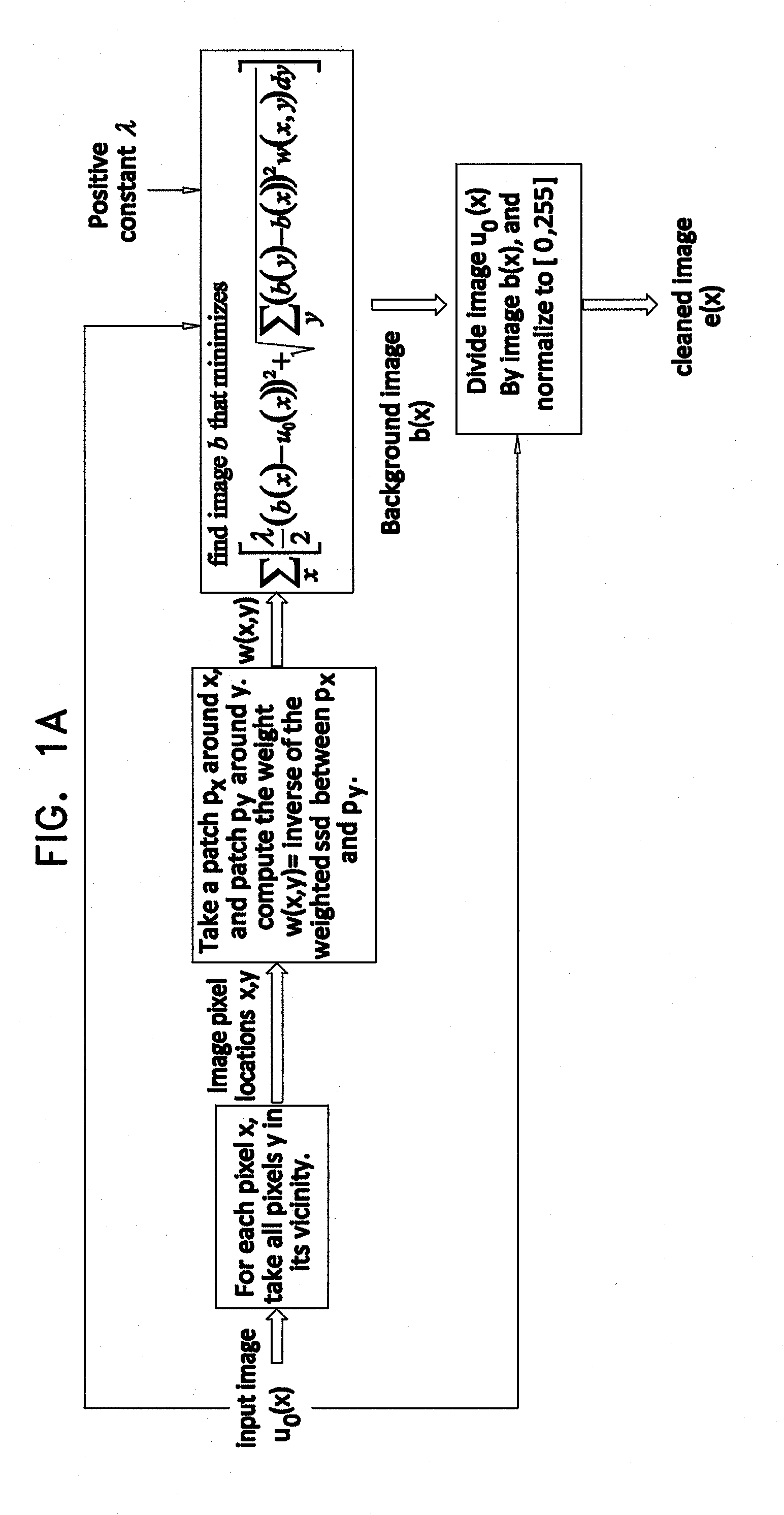
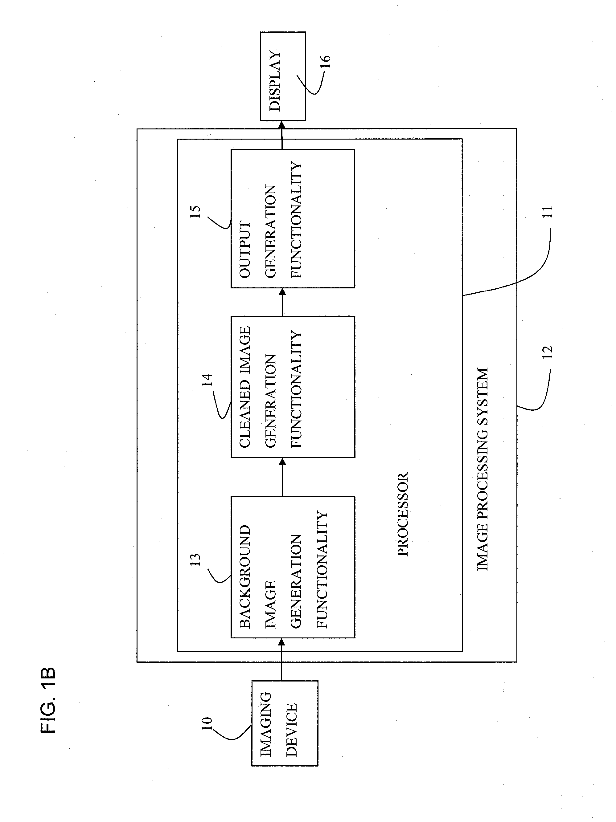
[0033]Applications of the present invention generally relate to medical image processing. Specifically, applications of the present invention relate to background cleaning in images of body lumens and body cavities.

BACKGROUND OF THE INVENTION

[0034]Vascular catheterizations, such as coronary catheterizations, are frequently-performed medical interventions. Such interventions are typically performed in order to diagnose the blood vessels for potential disease, and / or to treat diseased blood vessels. Typically, in order to facilitate visualization of blood vessels, the catheterization is performed under extraluminal imaging. Typically, and in order to highlight the vasculature during such imaging, a contrast agent is periodically injected into the applicable vasculature. The contrast agent typically remains in the vasculature only momentarily. During the time that the contrast agent is present in the applicable vasculature, the contrast agent typically hides, in full or in part, or obs...


PUM


Abstract

Apparatus and methods are described for use with an input angiogram image of a device inserted inside a portion of subject's body, the angiogram image being acquired in the presence of contrast agent. At least one processor (11) includes background-image-generation functionality (13) configured to generate a background image in which a relative value is assigned to a first pixel with respect to a second pixel, at least partially based upon relative values of surroundings of the first pixel and the surroundings of the second pixel in the input image. Cleaned-image-generation functionality (14) generates a cleaned image in which visibility of the radiopaque portions of the device is increased relative to the input image, by dividing the input image by the background image. Output-generation functionality (15) drives a display (16) to display an output based upon the cleaned image. Other applications are also described.

Description

CROSS REFERENCES TO RELATED APPLICATIONS[0001]The present application claims priority from U.S. Provisional Patent Application 61 / 457,866 to Barzelay, filed Jun. 23, 2011.[0002]The present application is related to International Patent Application PCT / IL2011 / 000612 (published as WO 12 / 014,212), entitled “Co-use of endoluminal data and extraluminal imaging,” filed Jul. 28, 2011, which:[0003](a) claims the benefit of:[0004]U.S. Provisional Patent Application 61 / 344,464, entitled “Co-use of endoluminal data and extraluminal imaging,” filed 29 Jul. 2010;[0005]U.S. Provisional Patent Application 61 / 344,875, entitled “Co-use of endoluminal data and extraluminal imaging,” filed 1 Nov. 2010;[0006]U.S. Provisional Patent Application 61 / 457,339, entitled “Co-use of endoluminal data and extraluminal imaging,” filed 3 Mar. 2011;[0007]U.S. Provisional Patent Application 61 / 457,455, entitled “Co-use of endoluminal data and extraluminal imaging,” filed 1 Apr. 2011;[0008]U.S. Provisional Patent App...

Claims


Application Information

Patent Timeline
IPC IPC(8): G06T5/00
CPCG06T5/001A61B6/481A61B6/504G06T5/009G06T5/50G06T2207/10116G06T2207/30101A61B6/52A61B6/5235A61B6/5258G06T2207/20221G06T2207/20212
Inventor BARZELAY, ZOHARCOHEN, RANTOLKOWSKY, DAVID
Owner SYNC RX
Why Eureka
Try Eureka