Clavicle suppression in radiographic images

a radiographic image and clavicle technology, applied in the field of radiographic imaging, can solve the problems of radiologist misinterpretation or overlooking tissue features of interest, difficult to remove rib features from the chest x-ray image without degrading, and higher false positive rates

Active Publication Date: 2014-05-22
CARESTREAM HEALTH INC
View PDF23 Cites 16 Cited by
  • Summary
  • Abstract
  • Description
  • Claims
  • Application Information

AI Technical Summary

Problems solved by technology

Even if rib structures are well-defined, however, it can be challenging to remove rib features from the chest x-ray image without degrading the underlying image content that can include lung tissue.
Poor performance in detecting and suppressing the ribs translates to higher rates of false positives (FPs) in diagnosing the lung tissue and can cause the radiologist to misinterpret or overlook tissue features of interest.
Suppression of the clavicle presents a particular challenge for image processing and it can be more difficult to accurately identify the clavicle due to its particular structure and conventional chest x-ray imaging practices.
In the x-ray image, the clavicle structure crosses the ribs and the intersection of the rib cage with the clavicle can readily confuse rib detection algorithms, leading to less than ideal rib segmentation and poor results in clavicle suppression.

Method used


Image

  • Clavicle suppression in radiographic images
  • Clavicle suppression in radiographic images
  • Clavicle suppression in radiographic images

Examples


Embodiment Construction

[0036]The following is a detailed description of the preferred embodiments of the invention, reference being made to the drawings in which the same reference numerals identify the same elements of structure in each of the several figures.

[0037]Reference is made to U.S. Provisional Patent Application No. 61 / 727,769 filed Nov. 19, 2012 entitled “CLAVICLE SUPPRESSION IN RADIOGRAPHIC IMAGES” in the names of Hui Zhao and Zhimin Huo and incorporated herein in its entirety.

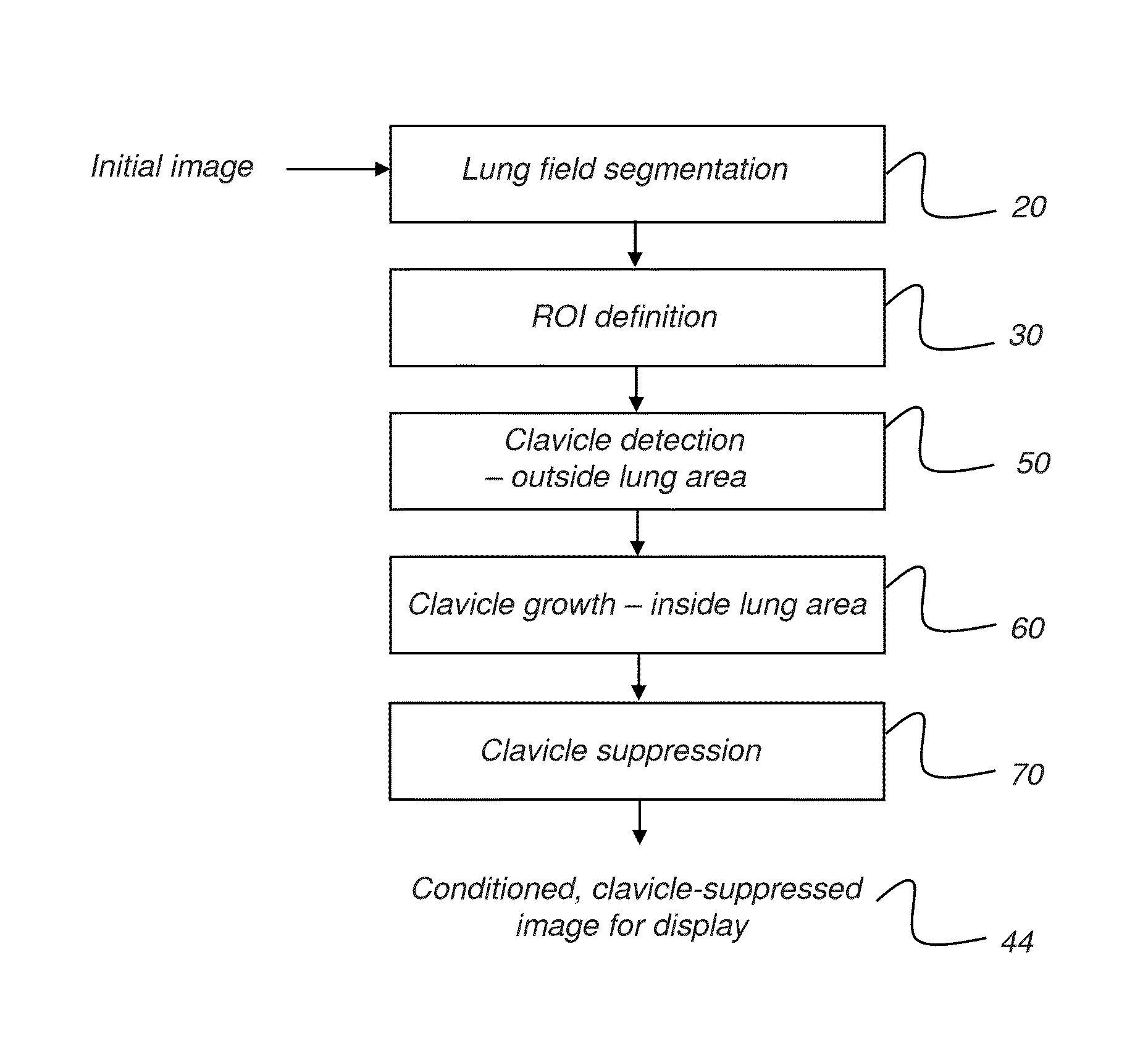
[0038]In the context of the present disclosure, the term “segmentation” has the broad meaning that is generally understood by those skilled in the image processing arts. The act of segmenting the image partitions the image content in some way so that one or more sets of pixels are grouped according to the feature(s) they represent. Thus, for a chest x-ray for example, lung segmentation defines those portions of the image that represent lung tissue for a patient.

[0039]The logic flow diagram of FIG. 1 shows a sequence for ...


PUM


Abstract

A method for clavicle suppression in a chest x-ray image. The method identifies the lung fields in the x-ray image and detects at least one portion of a clavicle ridge that lies outside the lung fields. Edges of a clavicle on each side of the detected clavicle ridge are detected, edge detection extended for the clavicle edges into the lung fields, and the clavicle defined within the x-ray image according to the edge detection. The clavicle is suppressed within the x-ray image to generate a clavicle-suppressed x-ray image and the clavicle-suppressed x-ray image is displayed, stored, or transmitted.

Description

CROSS REFERENCE TO RELATED APPLICATIONS[0001]Priority is claimed from U.S. Provisional Patent Application No. 61 / 727,769 filed Nov. 19, 2012 entitled “CLAVICLE SUPPRESSION IN RADIOGRAPHIC IMAGES” in the names of Hui Zhao and Zhimin Huo and incorporated herein in its entirety.FIELD OF THE INVENTION[0002]The present invention relates generally to the field of radiographic imaging and more particularly to methods for detecting and suppressing clavicle image content in chest x-ray images.BACKGROUND OF THE INVENTION[0003]The chest x-ray is useful for detecting a number of patient conditions and for imaging a range of skeletal and organ structures. Radiographic images of the chest are useful for detection of lung nodules and other features that indicate lung cancer and other pathologic structures and other life-threatening conditions. In clinical applications such as in the Intensive Care Unit (ICU), chest x-rays can have particular value for indicating pneumothorax as well as for tube / li...

Claims


Application Information

Patent Timeline
Patent Type & Authority Applications(United States)
IPC IPC(8): G06T7/00
CPCG06T7/0012G06T5/20G06T2207/10116G06T2207/20036G06T2207/20081G06T2207/20084G06T2207/30008G06T2207/30061G06T7/12G06T5/005
Inventor HUO, ZHIMINZHAO, HUI
Owner CARESTREAM HEALTH INC
Why Eureka
Try Eureka