Process for Devitalizing Soft-Tissue Engineered Medical Implants, and Devitalized Soft-Tissue Medical Implants Produced

a technology of soft tissue and engineered medical implants, which is applied in the field of devitalizing soft tissue engineered medical implants and devitalizing soft tissue medical implants produced, can solve the problems of limited success, inability to meet the needs of patients, and accumulation of residuals from various processing solutions, so as to achieve the effect of reducing processing times and conditions, improving production efficiency, and optimizing processing times and conditions

Inactive Publication Date: 2014-06-05
LIFENET HEALTH
View PDF1 Cites 12 Cited by
  • Summary
  • Abstract
  • Description
  • Claims
  • Application Information

AI Technical Summary

Benefits of technology

[0005]The inventive process described herein produces devitalized grafts including but not limited to ligaments, tendons, menisci, cartilage, skin, pericardium, dura mater, fascia, small and large intestine, placenta, veins, arteries, and heart valves. The methodology is advantageous over prior art techniques in that processing times and conditions have been optimized and reduced. In addition, the economics of production have been dramatically improved, resulting in large numbers of uniform, non-immunogenic grafts that repopulate with physiologically competent recipient cells. The grafts' collagenous matrix is substantially free from damage and retains mid-molecular weight cytoskeletal proteins for the purpose of acting as a chemoattractant to the infiltrating cells, such that those cells that infiltrate mature into the appropriate cell type. The grafts are also substantially free from contamination including, for example, free from infectious agents.
[0006]The invention embodies the use of one or more non-denaturing agents, for example, N-lauroyl sarcosinate, to solubilize the cellular components of the tissue and leave the matrix such that it is suitable for recellularization upon implantation. The invention is directed at a process for producing devitalized, soft-tissue implants including vascular grafts, veins, arteries, heart valves, tendons, ligaments, intervertebral discs, menisci and articular cartilage where processing times and conditions have been optimized to dramatically improve the economics of production as well as to produce a graftt with minimum damage to the matrix structure of the devitalized graft.

Problems solved by technology

The prior art processing methods are directed to decellularizing tissue grafts, i.e. removing all cellular elements, leaving a tissue matrix free from cellular elements, and are prohibitively time consuming, easily requiring numerous days, for example anywhere from eight to twenty-one days total processing time.
Such long processing times result in proteolytic degradation of the matrix structures of the processed tissues and may result in the accumulation of residuals from the various processing solutions.
While vascular prostheses are available for clinical use, they have met with limited success due to cellular and immunological complications, and the inability to remain patent and functional.
These problems are especially pronounced for small diameter prostheses, i.e. less than about 6 mm.
In addition, the methods used in the prior art dictate the use of large volumes of processing solutions which do not lend themselves to the production of acellular grafts on a commercializable scale.

Method used


Image

  • Process for Devitalizing Soft-Tissue Engineered Medical Implants, and Devitalized Soft-Tissue Medical Implants Produced
  • Process for Devitalizing Soft-Tissue Engineered Medical Implants, and Devitalized Soft-Tissue Medical Implants Produced
  • Process for Devitalizing Soft-Tissue Engineered Medical Implants, and Devitalized Soft-Tissue Medical Implants Produced

Examples


example 1

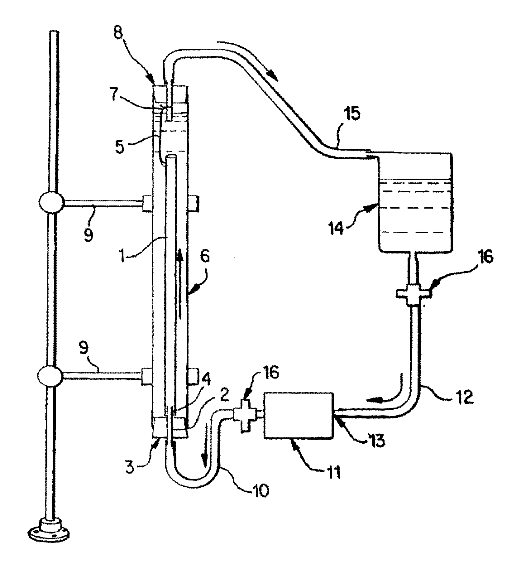
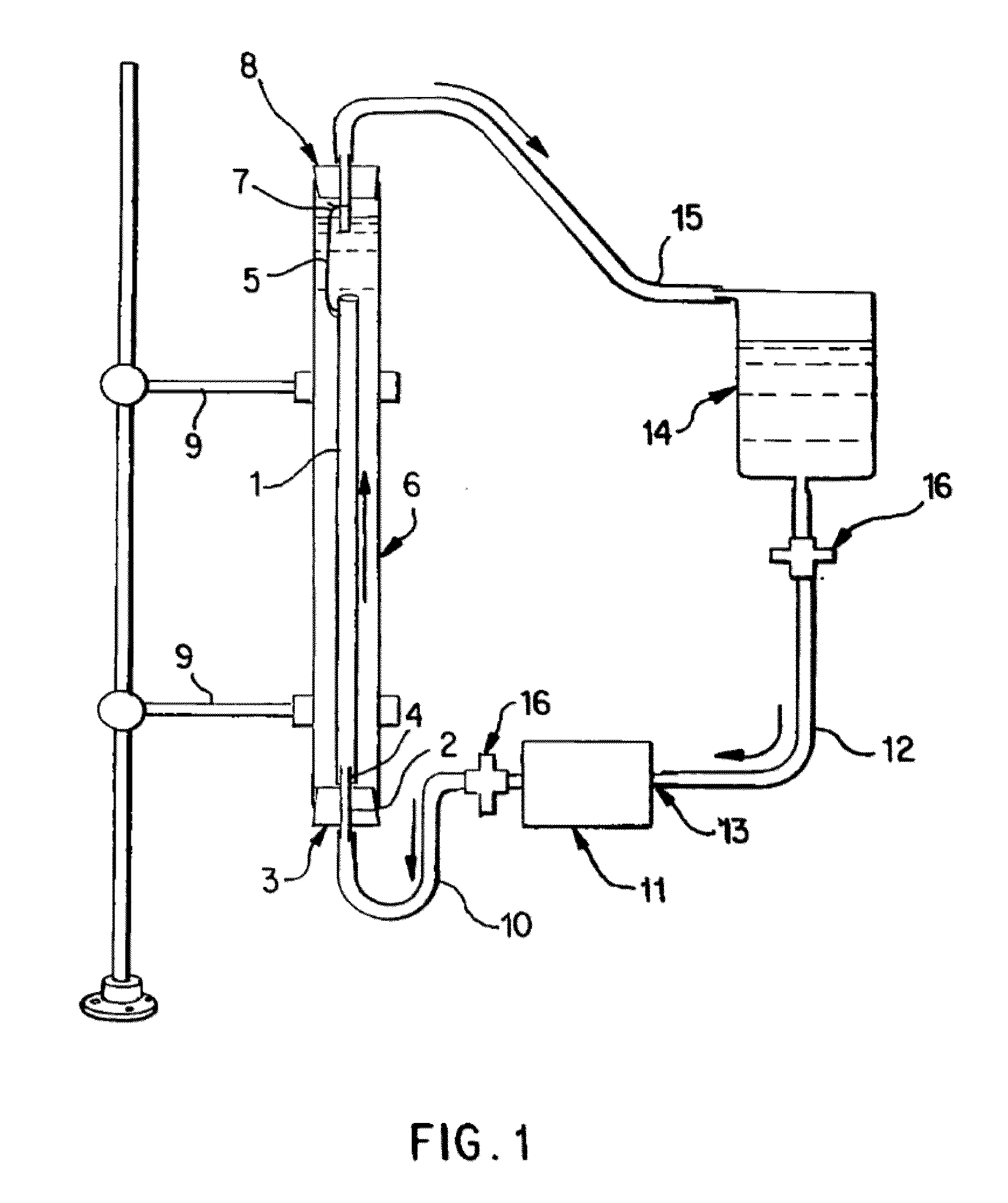
[0065]Saphenous vein tissues (two) from each leg of an acceptable human donor were carefully dissected under sterile conditions to remove all visible fat deposits and the side vessels were tied off using non-resorbable suture materials such that the ties did not occur in close proximity to the long run of the vessel. Sutures may restrict the devitalization process and the tissues under the sutures were removed following devitalization. For long vein grafts (40-60 cm) (FIG. 1), the distal ends of the veins were cannulated onto the ribbed attachment of the inlet ports and single sutures used to secure each vein. Additional suture lines were attached to the proximal ends of the veins. The veins were then removed from the dissecting solution (RPMI 1640, papaverine) and transferred to the processing vessel which had been temporarily inverted. The second suture line along with the vein was passed through the processing vessel and secured to a point on the outlet port end of the processing...

example 2

[0066]Saphenous vein tissues (two) from each leg of an acceptable human donor were carefully dissected under sterile conditions to remove all visible fat deposits and side vessels were tied off using nonresorbable suture materials such that the ties did not occur in close proximity to the long run of the vessel. Sutures may restrict the devitalization process and the tissues under the sutures were removed following devitalization. For long vein grafts (33 and 28 cm) (FIG. 1), the distal ends of the veins were cannulated onto the ribbed attachment of the inlet ports and single sutures used to secure each vein. Additional suture lines were attached to the proximal ends of the veins. The veins were removed from the dissecting solution (RPMI 1640, papaverin) and Polymixin B. Then the veins were cryopreserved according to current guidelines in RMPI 1640, 10% fetal calf serum, and 10% DMSO and control rate frozen at 1° C. / min and held in nitrogen vapor until devitalization. Prior to devit...

example 3

[0067]Internal mammary artery tissues (two) from an acceptable human donor were carefully dissected under sterile conditions to remove all visible fat deposits and side vessels were tied off using nonresorbable suture materials such that the ties did not occur in close proximity to the long run of the vessel. Sutures may restrict the devitalization process and the tissues under the sutures were removed following devitalization. For short artery grafts (11 and 8 cm) (FIG. 1), one end of each artery was cannulated onto the ribbed attachment of the inlet ports and single sutures used to secure each artery. The arteries were then removed from the dissecting solution (RPMI 1640 and papaverine) and one or more antibiotics and transferred to the processing vessel which had been temporarily inverted. Prior to closing the processing vessel, a portion of the extracting solution was gently added to the processing vessel and the inlet port, with attached artery, was then secured. At this point,...


PUM

PropertyMeasurementUnit
processing timeaaaaaaaaaa
diameteraaaaaaaaaa
thicknessaaaaaaaaaa

Abstract

The invention provides methodologies and apparatus for producing devitalized soft-tissue implants where the implant retains metabolically non-viable and / or reproductively non-viable cells, and preferably retains large molecular weight cytoplasmic proteins, such implants produced both in small quantities and in commercializable quantities. Such soft-tissue implants include vascular graft substitutes. A devitalized graft is produced by subjecting the tissue sample to an induced pressure mediated flow of an extracting solution, optionally followed by inducing a pressure mediated flow of a salt solution, then washing the tissue to produce the devitalized graft. The devitalized grafts produced are uniform and non-immunogenic. The inventive method allows for the production of multiple devitalized soft tissue implants, where processing time is significantly less than prior art processes and the number of implants produced per day is increased over prior art processes. In clinical use, the devitalized grafts produced exhibit significantly improved in long-term durability and function, and enhanced recellularization post-implantation.

Description

[0001]This invention is a continuation-in-part application of U.S. patent application Ser. No. 09 / 660,422, filed Sep. 12, 2000, which is now U.S. Pat. No. ______.FIELD OF THE INVENTION[0002]The invention is directed toward methodologies and apparatus for use in the preparation of devitalized soft tissue implants, i.e. essentially lacking in reproductively viable cells and / or metabolically viable cells, while preferably retaining reproductively non-viable cells and / or metabolically non-viable cells and / or large molecular weight cytoplasmic proteins including actin. These devitalized, soft-tissue implants, are produced in small and commercializable quantities and include vascular and musculoskeletal graft substitutes. These implants may be derived from tissue-engineered soft tissues, tissue products derived from animal or human donors that contain cells which contain or are devoid of valve structures useful in directing the flow of fluids through tubular vascular prostheses, and / or co...

Claims


Application Information

Patent Timeline
Patent Type & Authority Applications(United States)
IPC IPC(8): A01N1/02A01N1/00A61K35/12A61L27/36C12N5/00C12N5/08
CPCA01N1/00A01N1/02A01N1/0226A01N1/0247A61K35/12A61L27/3604A61L27/3683A61L27/3691C12N5/00C12N2509/00
Inventor WOLFINBARGER, LLOYDLANGE, PERRYLINTHURST-JONES, ALYCEMOORE, ERICNOLF, BARRY
Owner LIFENET HEALTH
Why Eureka
Try Eureka