
Method for efficient digital subtraction angiography

a digital subtraction and angiography technology, applied in image data processing, diagnostics, applications, etc., can solve the problems of inefficiency, unfavorable subtraction process, and many drawbacks,

Active Publication Date: 2014-09-18
PHILIPS MEDICAL SYST TECH
View PDF28 Cites 22 Cited by
  • Summary
  • Abstract
  • Description
  • Claims
  • Application Information

AI Technical Summary

Benefits of technology

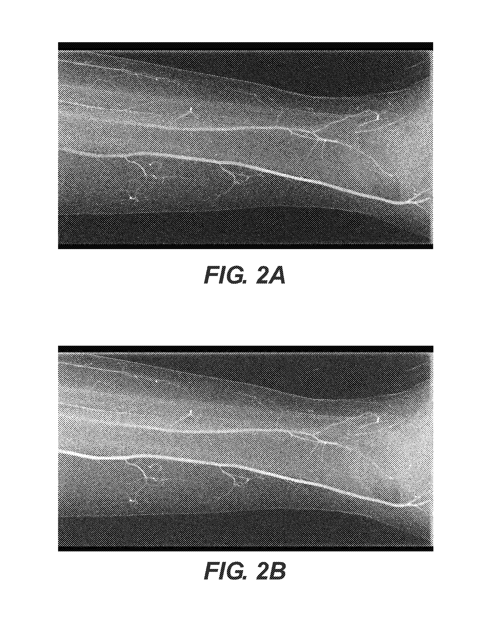
The present invention provides a system and method for efficiently performing DSA (digital subtraction angiography) without the need for a non-contrast initial or "mask" image. Instead, multiple contrast enhanced images are obtained and optionally registered with each other using a rigid or non-rigid registration method. The images are then subtracted from each other to obtain the desired results. The process of obtaining the results is efficient and does not require a separate mask image. The invention also provides a cine view of the results and noise reduction algorithms to improve the quality of the results. The technical effects of the invention include improved efficiency and accuracy of DSA, reduced time consumption, and improved image quality.

Problems solved by technology

The background art describes methods to perform DSA, which unfortunately suffer from a number of drawbacks and inefficiencies.
However, if any movement occurs between obtaining the non-enhanced image and the enhanced image, these images will not be in registration and so the subtraction process will not be performed correctly.

Method used


Image

  • Method for efficient digital subtraction angiography
  • Method for efficient digital subtraction angiography
  • Method for efficient digital subtraction angiography

Examples


Embodiment Construction

[0016]At least some embodiments of the present invention are now described with regard to the following illustrations and accompanying description, which are not intended to be limiting in any way.

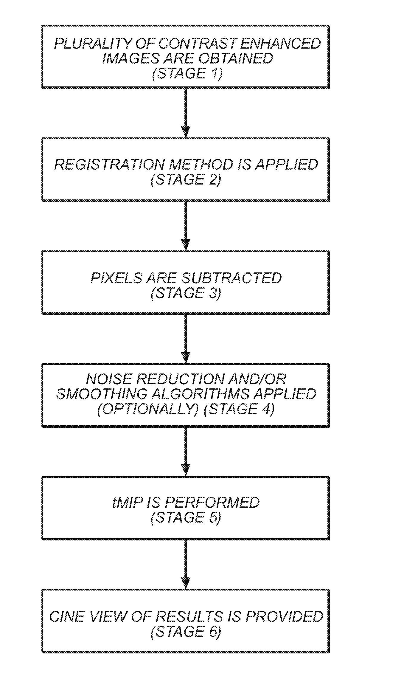
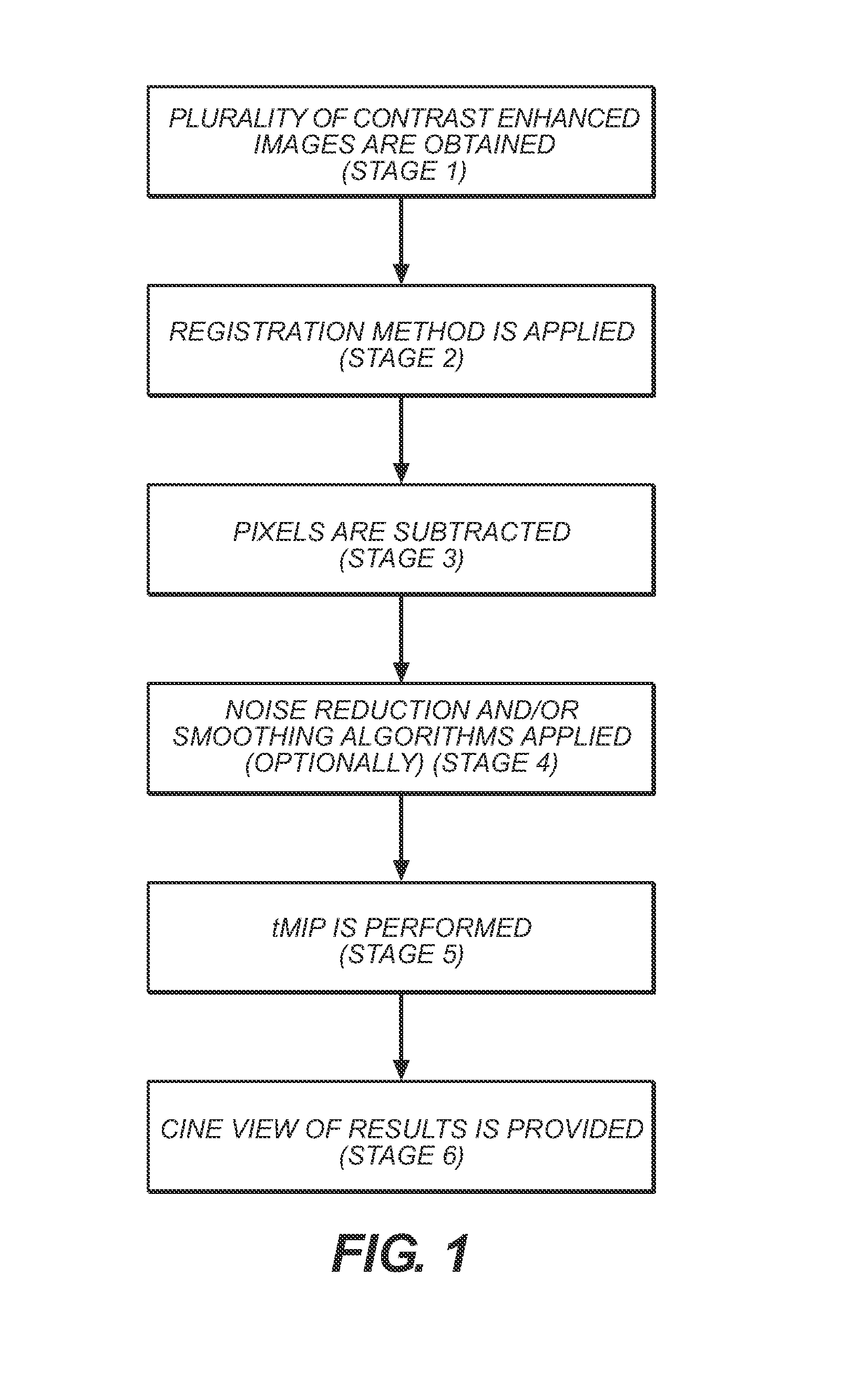
[0017]Referring now to the drawings, FIG. 1 shows an exemplary, illustrative method for efficiently performing DSA (digital subtraction angiography) according to at least some embodiments of the present invention. In stage 1, a plurality of contrast enhanced images are obtained, such that at least two but preferably at least three images are obtained and more preferably 10 images are obtained (or even more). Next, in stage 2, optionally and preferably a registration is performed between these images, whether rigid or non-rigid. Optionally and preferably registration is performed by using a known registration method, non-limiting examples of which are described with regard to “Algorithms for radiological image registration and their clinical application” by Hawkes et al (J. Anat. (1998) 193...


PUM


Abstract

A system and method for performing DSA (digital subtraction angiography), which does not require a non-enhanced or “mask” image to be obtained.

Description

FIELD OF THE INVENTION[0001]The present invention relates to a system and method for an alternative method of performing efficient digital subtraction angiography and particularly, but not exclusively, to automatic efficient digital subtraction angiography in image data of bodily tissue and / or object feature analysis in image data from non-biological subjects.BACKGROUND OF THE INVENTION[0002]Angiography refers generally to the capture and representation of blood vessels or vasculature of the human body by means of X-ray imaging, i.e., X-ray vascular imaging. X-ray diagnostic imaging systems may be used for angiographic imaging procedures such as digital subtraction angiography (DSA), and live fluoroscopic roadmapping. As currently practiced in the art, digital subtraction angiography or DSA is an imaging method used for visualizing blood vessels inside a patient's body that includes injecting a contrast medium bolus that is substantially opaque to X-rays into the blood vessels or va...

Claims


Application Information

Patent Timeline
Patent Type & Authority Applications(United States)
IPC IPC(8): G06T7/00
CPCG06T7/0012G06T5/50G06T2207/20224G06T2207/30101A61B6/481A61B6/486A61B6/504G06T7/30G06T5/70
Inventor SHREIBER, REUVEN R.ENGELHARD, GUY E.
Owner PHILIPS MEDICAL SYST TECH
Learn More