Method for localization of an epileptic focus in neuroimaging

a neuroimaging and epileptic focus technology, applied in the field of neuroimaging epileptic focus localization, can solve the problems of too many potential focus areas, difficult interpretation of difference images acquired according to current methods, etc., and achieve the effect of reducing the amount of distracting information and facilitating interpretation

Active Publication Date: 2014-09-18
SIEMENS MEDICAL SOLUTIONS USA INC
View PDF0 Cites 10 Cited by
  • Summary
  • Abstract
  • Description
  • Claims
  • Application Information

AI Technical Summary

Benefits of technology

[0019]The present invention accordingly addresses the above issues and provides improved methods for identifying candidate foci in neuroimaging by reducing the amount of distracting information with the aim to facilitate interpretation.

Problems solved by technology

However, 25-30% of people with epilepsy do not have seizure control even with the best available medications.
However, difference images acquired according to current methods can be difficult to interpret as they may show too many potential areas of high variation, most of which are in fact irrelevant, and distracting.
Few techniques exist to facilitate the location of epileptic foci.
However, this has often been found to identify too many potential focus areas.

Method used


Image

  • Method for localization of an epileptic focus in neuroimaging
  • Method for localization of an epileptic focus in neuroimaging

Examples


Embodiment Construction

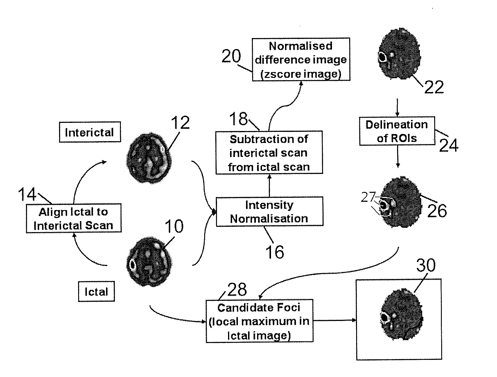
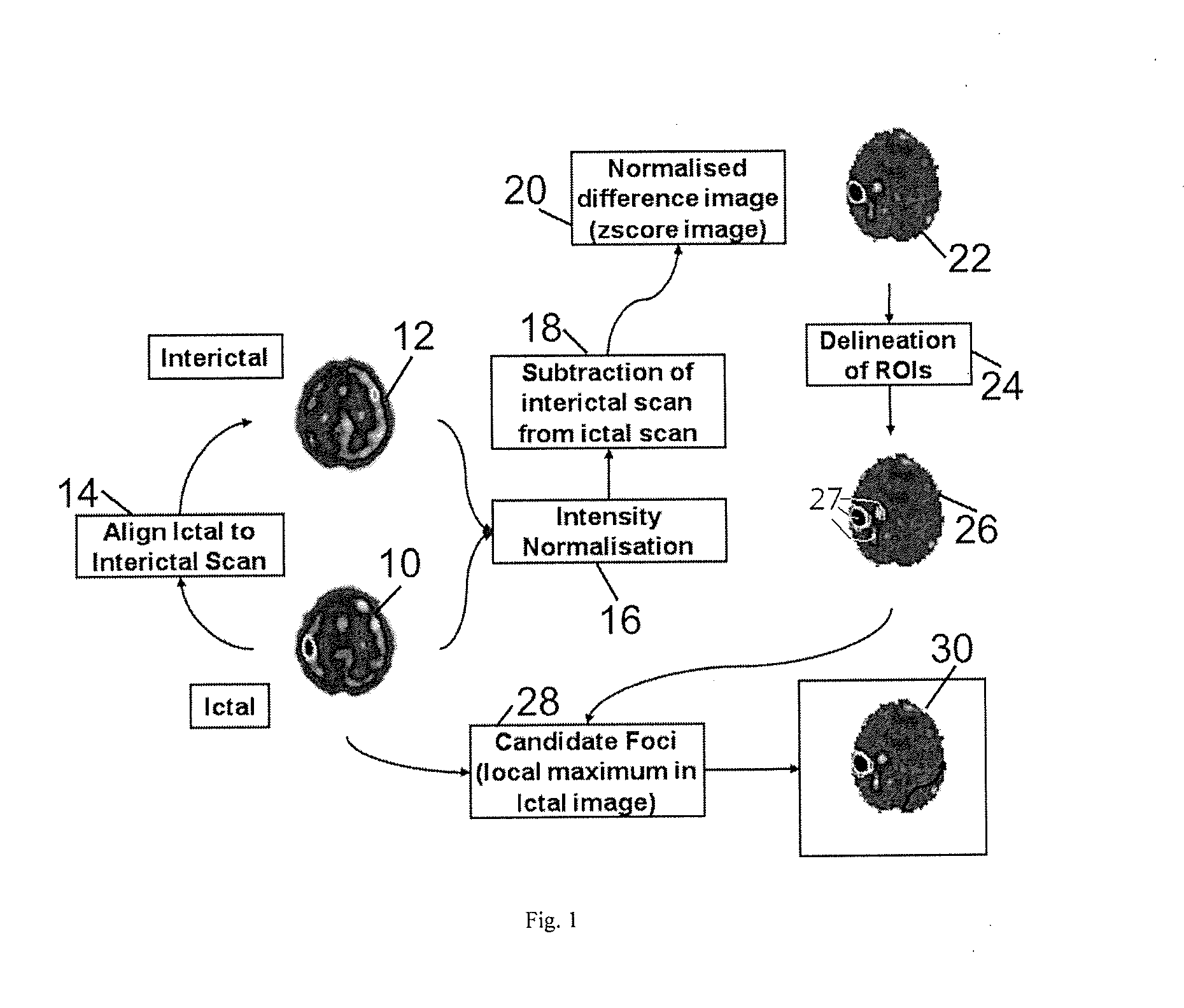
[0022]The present invention provides a method for identifying candidate foci, in which no comparisons are made to a normal database, but the necessary information is extracted directly from an ictal-interictal difference image, itself derived as defined in the SISCOM method discussed above.

[0023]According to a method of the present invention, the process proceeds as in the conventional method described above as far as normalizing the difference image to a Z-score image and applying a threshold corresponding to number of standard deviations. By applying such a threshold, all potential focus regions in the difference image are identified.

[0024]As discussed above, this may result in identifying many potential focus regions. To reduce the amount of distracting information produced in the method so far, the present invention provides further steps in which the difference image is analyzed, and the intervention (ictal) image is compared to the analyzed difference image to confirm or rejec...


PUM


Abstract

In a method for localizing a candidate foci in neuroimaging, two images are acquired, one being a baseline (interictal) image and another being an intervention (ictal) image. The two images are aligned and the intensities of the images are normalized. A difference image is calculated by subtracting the baseline (interictal) image from the intervention (ictal) image. The difference image is normalized and regions of interest are selected as candidate foci.

Description

BACKGROUND OF THE INVENTION[0001]1. Field of the Invention[0002]The present invention concerns a method for localization of an epileptic focus in neuroimaging.[0003]2. Description of the Prior Art[0004]The following definitions, acronyms, and abbreviations are used herein:[0005]ECD Ethyldysteinatdimers[0006]HMPAO Hexamethylpropyleneamine Oxime[0007]MRI Magnetic Resonance Imaging[0008]PET Positron Emission Tomography[0009]SISCOM Subtraction Ictal SPECT Co-registered to MRI[0010]SPECT Single-Photon Emission Computed Tomography[0011]Epilepsy is a brain disorder in which a person has repeated seizures over time. Approximately 1% of the UK population is affected by epilepsy which is usually controlled (but not cured) with medication. However, 25-30% of people with epilepsy do not have seizure control even with the best available medications.[0012]Surgery may be considered in difficult cases, where a required evaluation will include neurological examination, routine EEG, Long-term video-E...

Claims


Application Information

Patent Timeline
Patent Type & Authority Applications(United States)
IPC IPC(8): G06T7/00
CPCG06T7/0012G06T7/0016G06T2207/10088G06T2207/10104G06T2207/10108G06T2207/30016A61B6/037A61B6/5217G06T7/254G06T5/50A61B5/4094
Inventor DECLERCK, JEROMESADDI, KINDA ANNA
Owner SIEMENS MEDICAL SOLUTIONS USA INC
Why Eureka
Try Eureka