Functional magnetic resonance imaging biomarker of neural abnormality

a functional magnetic resonance imaging and biomarker technology, applied in the field of functional magnetic resonance imaging, can solve the problems of not being able to objectively detect cognitive impairments due to disease or injury by conventional standard tests, and the medical industry has yet to establish a reliable solution for detecting and tracking the long-term consequences of repetitive mtbi in living subjects

Inactive Publication Date: 2014-09-18
HUBBARD DAVID R
View PDF9 Cites 18 Cited by
  • Summary
  • Abstract
  • Description
  • Claims
  • Application Information

AI Technical Summary

Benefits of technology

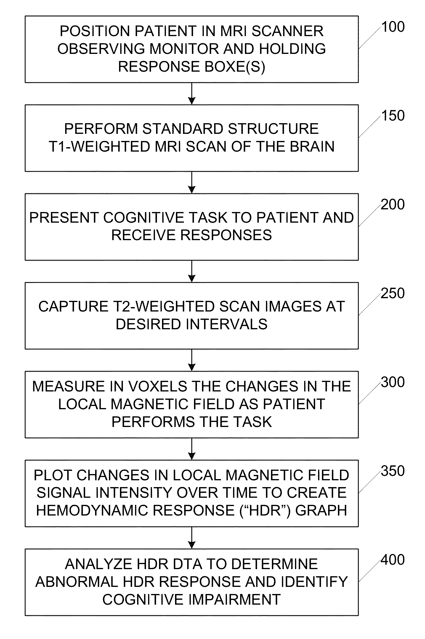
[0011]Functional magnetic resonance imaging (“fMRI”) is used to capture complete scans of the brain during the HDR process, which typically takes about 15-60 seconds per stimulus event. The result of fMRI is a series of scans of the subject over time that show what region of the brain was active during the HDR. A single scan includes a full set of slices that cover the brain of the subject. Each slice is a separate image and collectively the slices comprise a three dimensional image of the brain of the subject. Typically, an image is taken every 1-4 seconds. In this fashion, fMRI is used to identify the region of the brain that is active for a particular cognitive task and any number of cognitive tasks can be presented to the subject (e.g., in the range of 5-60 cognitive tasks). fMRI allows for strong functional-anatomical identification and fMRI scans have been used to locate important brain regions, such as language regions. fMRI scans have also been used in lie detection, because the attempt to deceive causes intense activity in several distinct brain areas. Data resulting from fMRI has been found to be robust and reproducible, and fMRI has become a helpful tool of cognitive neuroscience researchers.

Problems solved by technology

Finally, NFL alumni may also be at increased risk of developing chronic traumatic encephalopathy (“CTE”), a progressive tauopathy that is characterized by behavioural changes, memory impairments and Parkinsonism.
Despite the critical importance of these revelations regarding the effects of mTBI, the medical industry has yet to establish a reliable solution for detecting and tracking the long-term consequences of repetitive mTBI in living subjects.
For example, one recent study reported that NFL alumni were impaired on global measures of cognitive function while an earlier investigation failed to find any such impairment.
Additionally, cognitive impairments due to disease or injury are often not objectively detectable by conventional standard tests, such as a clinical neurological exam or anatomical imaging scanning.
Standard neuropsychological tests, whether paper-and-pencil or computerized, are typically susceptible to poor effort or malingering.
It should be understood that fMRI is a technically challenging procedure, and analysis and interpretation of fMRI data is complicated.
As a result, fMRI results are at risk of overinterpretation or misuse by those without adequate training.
This is a key issue as fMRI becomes a more widely used clinical tool to assess the functional impact of structural abnormalities from trauma or other lesions.

Method used


Image

  • Functional magnetic resonance imaging biomarker of neural abnormality
  • Functional magnetic resonance imaging biomarker of neural abnormality
  • Functional magnetic resonance imaging biomarker of neural abnormality

Examples


example study

[0047]A study was recently conducted in San Diego, Calif. to analyze the use of fMRI to identify neural abnormality in subjects having multiple mTBIs.

[0048]Participants: The FANTAB normative database was used for this study. Sixty healthy volunteers from the FANTAB database were recruited from the San Diego area for this study. All volunteers are right-handed college graduates, fluent in English, with normal or corrected-to-normal eyesight, normal hearing, and no history of neurological or psychiatric illnesses. These individuals were selected to cover a range of ages, between 20 and 60 years old. In the current study, an age-matched subset of twenty volunteers was compared to the NFL group (mean age controls=53 years old, NFL alumni=54 years old). Thirteen retired American professional football players participated in the neuroimaging study. They had no diagnosis of neurological or psychiatric illness. A further two retired players volunteered but could not be included in the neuro...


PUM


Abstract

Systems and methods for application of fMRI as a biomarker of neural abnormalities are presented. A subject's activations of brain regions that occur while performing cognitive tasks in an fMRI scanning apparatus are measured. The resulting measurements are compared to the activations of brain regions that occur during each cognitive task for a control group. The comparison to the performance and brain activations of normal subjects determine the presence of cortical compensatory mechanisms that indicate neural abnormalities in the subject.

Description

RELATED APPLICATION[0001]The present application claims priority to U.S. provisional patent appl. No. 61 / 780,030 filed 13 Mar. 2013 and also claims priority to U.S. provisional patent appl. No. 61 / 909,631 filed 27 Nov. 2013, each of which is incorporated herein by reference in its entirety.BACKGROUND[0002]1. Field of the Invention[0003]The present invention is generally related to functional magnetic resonance imaging (“fMRI”) and is more specifically related to using fMRI as a biomarker of neural abnormality.[0004]2. Related Art[0005]Recent research has raised concerns regarding the prevalence of mild traumatic brain injuries (“mTBI”) and the potential for associated long-term neurological damage as a consequence of repetitive concussive and sub-concussive injuries. The Center for Disease Control and Prevention estimates that up to 3.8 million sports related concussions occur annually in the United States alone. A vast number of additional non-sports related concussions also occur ...

Claims


Application Information

Patent Timeline
Patent Type & Authority Applications(United States)
IPC IPC(8): G01R33/48
CPCG01R33/4806A61B5/055A61B5/4064
Inventor HUBBARD, DAVID R.
Owner HUBBARD DAVID R
Why Eureka
Try Eureka