Magnetic immuno digital PCR assay

Active Publication Date: 2014-11-13
BIO RAD LAB INC
View PDF0 Cites 7 Cited by
  • Summary
  • Abstract
  • Description
  • Claims
  • Application Information

AI Technical Summary

Benefits of technology

[0042]In still another aspect, kits for detecting an antigen or an antibody analyte in a sample are provided. In some embodiments, the kit comprises one or more affinity agents as described herein. In some embodiments, the one or more affinity agents are selected from the group consisting of antibodies, aptamers, and non-antibody protein scaffolds. In some embodiments, the affinity agent is an antibody.
[0043]Unless defined otherwise, technical and scientific terms used herein have the same meaning as commonly understood by a person of ordinary skill in the art. See, e.g., Lackie, DICTIONARY OF CELL AND MOLECULAR BIOLOGY, Elsevier (4th ed. 2007); Sambrook et al., MOLECULAR C

Problems solved by technology

However, the limits of detection and precision of quantification that c

Method used


Image

  • Magnetic immuno digital PCR assay
  • Magnetic immuno digital PCR assay
  • Magnetic immuno digital PCR assay

Examples


Example

Example 1

Scheme 1 for Detecting an Antigen

[0150]This example demonstrates detecting an antigen of interest using two primary affinity agents (e.g., antibodies), each of which specifically binds to the antigen. A sample is contacted with primary antibody A and primary antibody B. Antibody B has a magnetic bead covalently attached. Following incubation with antibodies A and B, the sample is contacted with a secondary antibody C that is labeled with a tagged nucleic acid (“TNA”). The antigen-primary antibody-secondary antibody-TNA complex is purified from free primary antibody A and secondary antibody C by magnetic separation with washing, then the purified complex is resuspended and captured in droplets. Droplet digital PCR is performed to detect the presence of the TNA label and / or quantify the amount of TNA label in each droplet. Detection and / or quantification of the antigen is then determined by mathematical conversion of the signal from the label to the number of antigens. See FI...

Example

Example 2

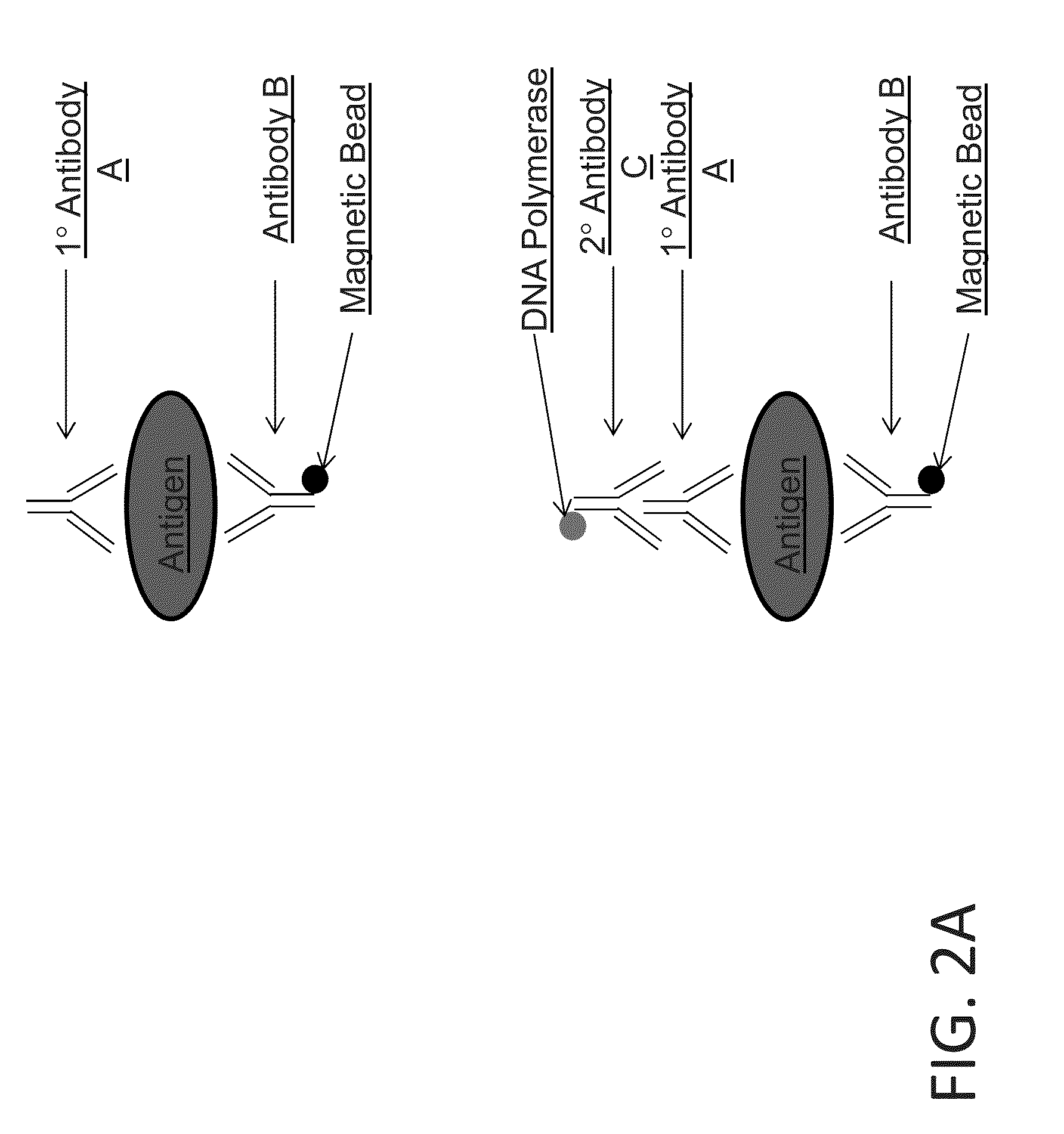
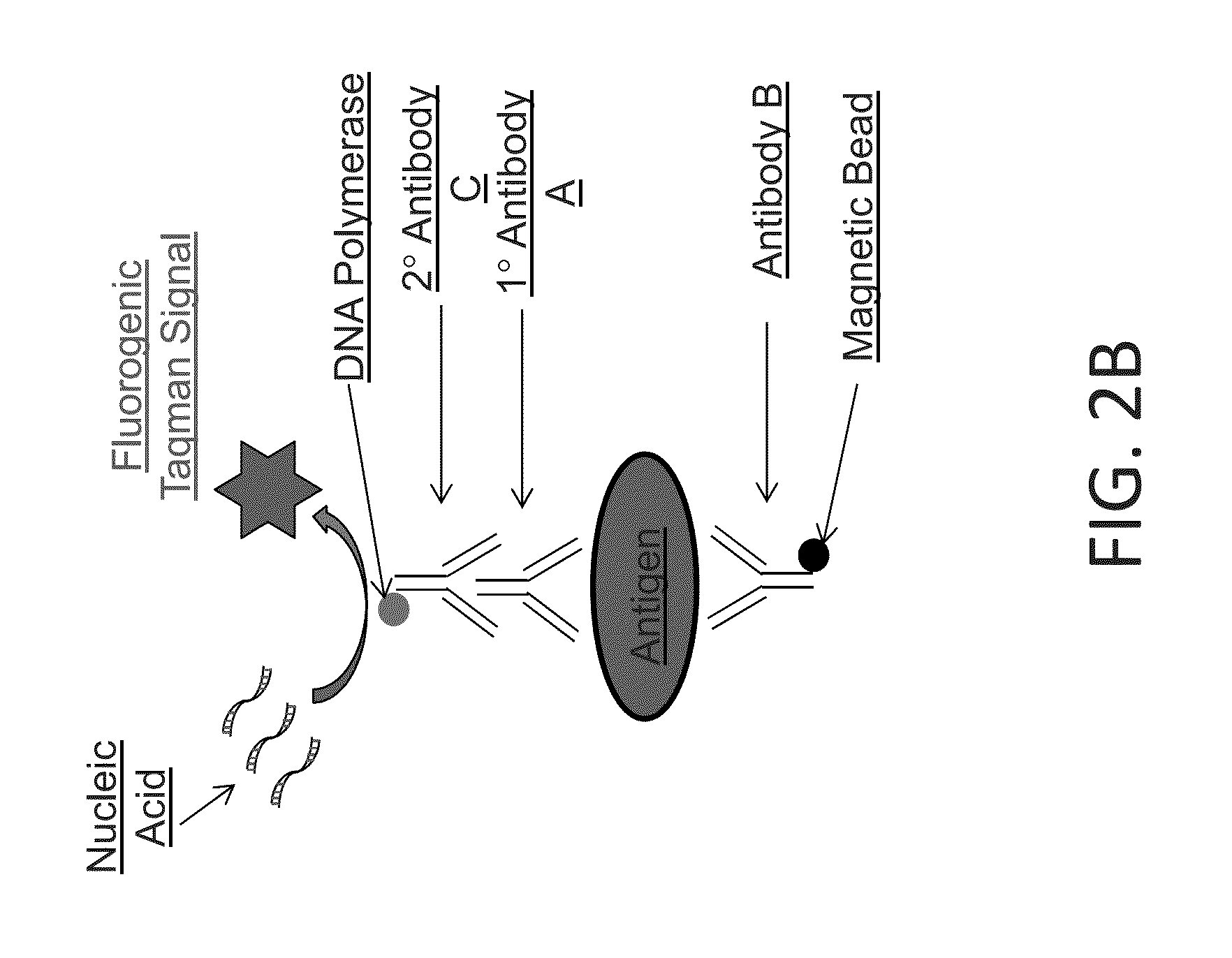
Scheme 2 for Detecting an Antigen

[0151]This example demonstrates detecting an antigen using two primary affinity agents (e.g., antibodies), each of which specifically binds to the antigen. In this scheme, an enzyme-linked secondary antibody is added that converts a substrate into a detectable form for detecting the presence of the antigen. A sample is contacted with primary antibody A and primary antibody B. Antibody B has a magnetic bead covalently attached. Following incubation with antibodies A and B, the sample is contacted with a secondary antibody C that binds specifically to antibody A. The secondary antibody C is coupled to a DNA polymerase such as Taq, Fast Start Taq, or some other polymerase (e.g., a mutagenized or variant DNA polymerase with desired properties for the application) or to another enzyme (FIG. 2A). The antigen-primary antibody-secondary antibody-DNA polymerase complex is purified from free primary and secondary antibodies by magnetic separation with...

Example

Example 3

Scheme 3 for Detecting an Antigen

[0152]This example demonstrates detecting an antigen of interest using two primary affinity agents (e.g., antibodies), each of which specifically binds to the antigen. A sample is contacted with primary antibody A and primary antibody B. Antibody B has a magnetic bead covalently attached. Following incubation with antibodies A and B, the sample is contacted with a secondary antibody C that is labeled with a fluorophore. The antigen-primary antibody-secondary antibody-fluorophore complex is purified from free primary antibody A and secondary antibody C by magnetic separation with washing, then the purified complex is resuspended and captured in droplets. The fluorescent signal generated by the fluorophore label is detected according to any method described herein. Detection and / or quantification of the antigen is then determined by mathematical conversion of the signal from the label to the number of antigens. See FIG. 4.


PUM

PropertyMeasurementUnit
Magnetismaaaaaaaaaa
Affinityaaaaaaaaaa

Abstract

The present invention provides methods of detecting an antigen in a sample comprising contacting the sample with affinity agents to form an antigen-affinity agent-label complex, separating the antigen-affinity agent-label complex from uncomplexed components, partitioning label from the separated sample into multiple partitions, and detecting the presence of the label in one or more partitions. Compositions and kits for detecting an antigen in a sample are also provided.

Description

CROSS-REFERENCE TO RELATED APPLICATIONS[0001]The present application claims priority to U.S. Provisional Application No. 61 / 821,530, filed May 9, 2013, and to U.S. Provisional Application No. 61 / 837,533, filed Jun. 20, 2013, each of which is incorporated by reference herein for all purposes.BACKGROUND OF THE INVENTION[0002]The quantification of antigens in a sample can provide useful information for a number of clinical applications. One method for detecting and quantifying antigens is by enzyme-linked immunosorbent assay (ELISA). However, the limits of detection and precision of quantification that can be achieved with this assay are not sufficient for many applications.[0003]Thus, there remains a need for methods of detecting and quantifying antigens that offer improved precision of quantification and low end sensitivity.BRIEF SUMMARY OF THE INVENTION[0004]In one aspect, methods of detecting an antigen in a sample are provided. In some embodiments, the method comprises:[0005]conta...

Claims


Application Information

Patent Timeline
IPC IPC(8): C12Q1/68
CPCC12Q1/6804C12Q2537/159C12Q2561/101C12Q2563/143C12Q2563/149
Inventor HODGES, SHAWNHEREDIA, NICKPETERSEN, JONATHAN
Owner BIO RAD LAB INC
Why Eureka
Try Eureka