Dental image display device, dental surgical operation device, and dental image display method

a technology of dental surgical operation and image display, which is applied in the direction of teeth capping, teeth nerve/root treatment implements, instruments, etc., can solve the problems of root canal orifice orifice orifice orifice breaking, etc., and achieve the effect of accurately grasping the position of the root canal orifice inside the tooth and avoiding fractures

Active Publication Date: 2014-11-20
MORITA MFG CO LTD
View PDF45 Cites 69 Cited by
  • Summary
  • Abstract
  • Description
  • Claims
  • Application Information

AI Technical Summary

Benefits of technology

[0013]One or more embodiments of the present invention provide a dental image display device, a dental surgical operation device, and

Problems solved by technology

For example, it is difficult to accurately grasp the position of the root canal orifice inside the tooth while checking an image of the surface of the tooth captured by the optical camera or the microsco

Method used


Image

  • Dental image display device, dental surgical operation device, and dental image display method
  • Dental image display device, dental surgical operation device, and dental image display method
  • Dental image display device, dental surgical operation device, and dental image display method

Examples


Embodiment Construction

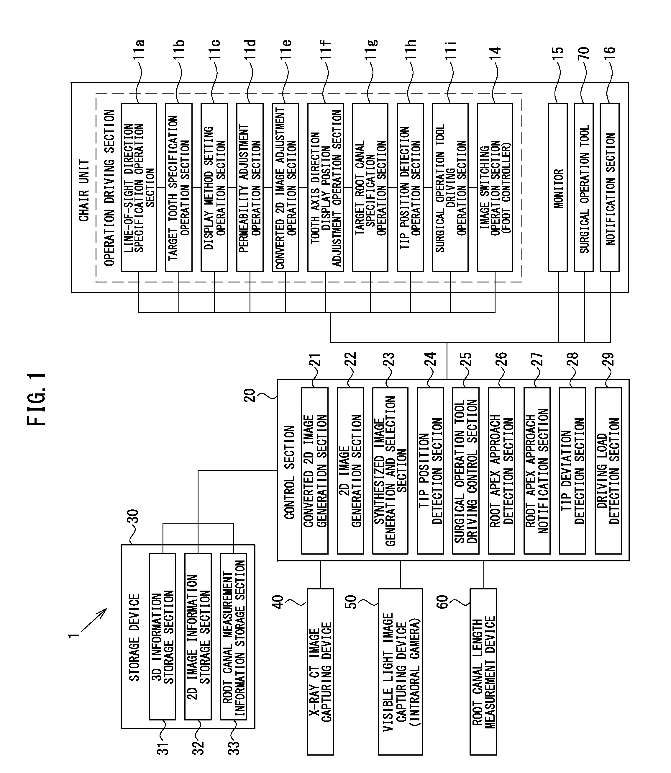
[0094]Hereinafter, a medical care system 1 according to one or more embodiments of the present invention will be described with reference to FIG. 1 through FIG. 19.

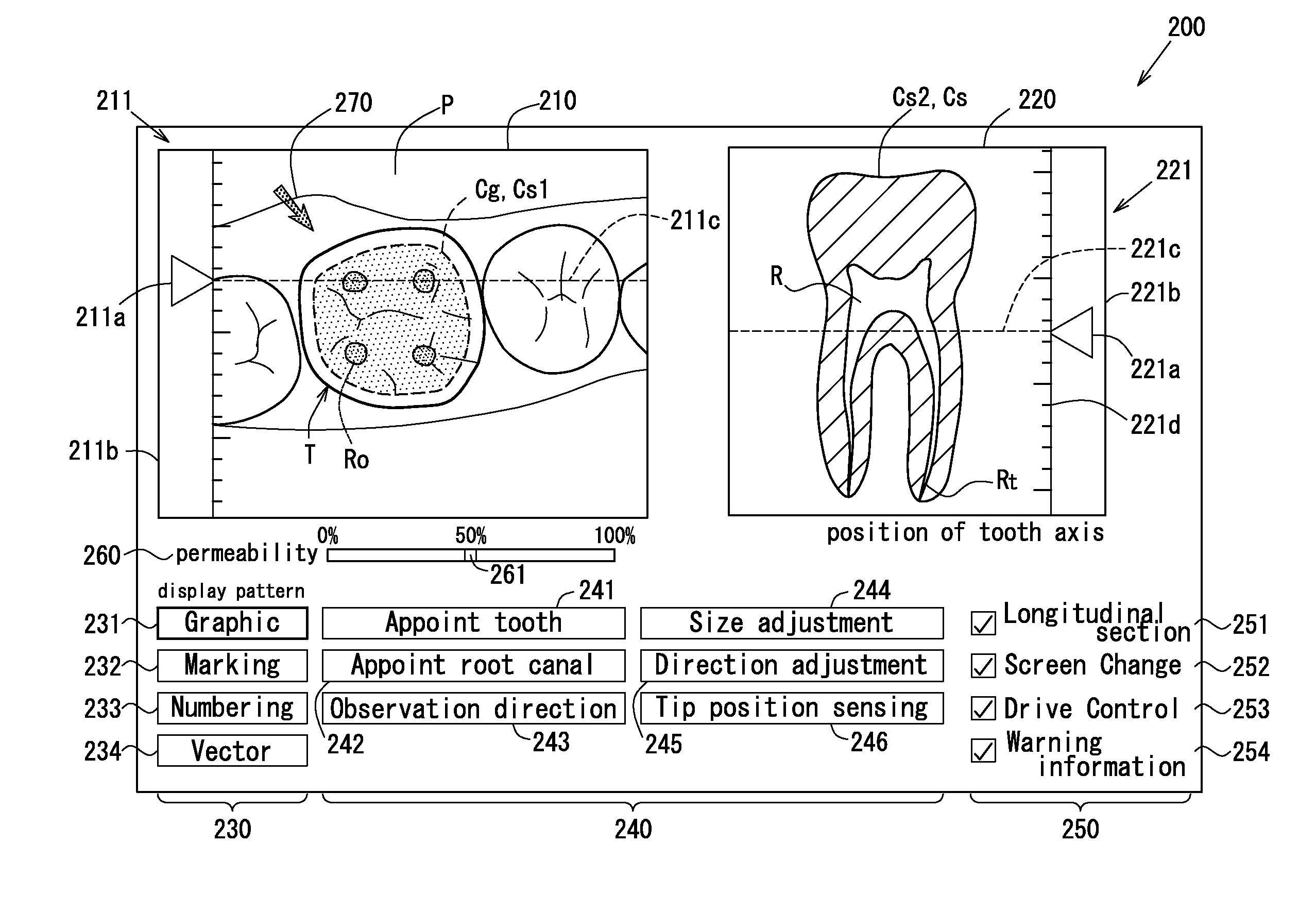
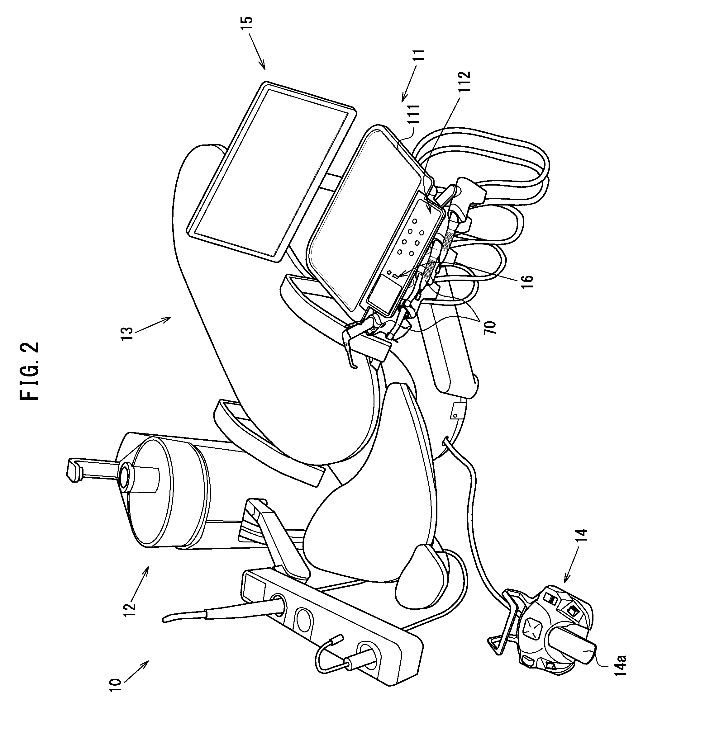
[0095]FIG. 1 is a block diagram of the medical care system 1, and FIG. 2 is a schematic isometric view showing a structure of a chair unit 10. FIG. 3 is a schematic isometric view of an X-ray CT image capturing device 40. FIG. 4 is a schematic view of an intraoral camera 50. FIG. 5 is a schematic isometric view of a root canal length measurement device 60. FIG. 6 is schematic isometric view of a surgical operation tool 70. FIG. 7 shows an image overlapping display screen 200.

[0096]FIG. 8 through FIG. 10 are flowcharts showing a dental care process. FIGS. 11A, 11B, and 11C show a method for adjusting a converted two-dimensional image. FIG. 12 shows marking overlapping display. FIGS. 13A, 13B, 13C, and 13D show overlapping display patterns. FIGS. 14A, 14B, and 14C show a method for adjusting permeability. FIGS. 15A and 15B ...


PUM


Abstract

According to a dental image display device, a dental surgical operation device, and a dental image display method, a 2D captured image of a tooth as a surgical operation target that is captured with visible light in an articulation face direction, and an articulation face direction converted 2D image obtained as a result of displaying 3D information, which is acquired on the tooth and includes information on a root canal inside the tooth, two-dimensionally on a predetermined plane in the articulation face direction, are displayed in correspondence with each other on a monitor.

Description

CROSS-REFERENCE TO RELATED APPLICATIONS[0001]The present application claims benefit of priority under 35 U.S.C. §119 to Japanese Patent Application No. 2013-043707 filed on Mar. 6, 2013. The contents of the priority applications are incorporated by reference in their entirety.BACKGROUND OF THE INVENTION[0002]1. Field of the Invention[0003]The present invention relates to a dental image display device, a dental surgical operation device, and a dental image display method for displaying a two-dimensional image of a tooth captured with visible light in an articulation face direction by an optical camera and also displaying two-dimensional display information generated based on three-dimensional information which is captured by an X-ray CT image capturing device or MRI and includes information on a root canal inside the tooth.[0004]2. Description of the Prior Art[0005]Conventionally, in the fields of dental care and the like, an image of a tooth is captured as follows for performing a s...

Claims


Application Information

Patent Timeline
IPC IPC(8): A61C5/02G06T15/08
CPCA61C5/025G06T2215/16G06T2200/04G06T15/08A61B6/032A61B6/14A61B6/463A61B6/5247A61B5/0088A61B5/0066A61B5/055A61B5/4542A61B5/4547A61C5/44
Inventor FLEER, JURGEN RICHARDNAKAI, TERUJISUZUKI, MASAKAZUSADAKANE, TOMOYUKI
Owner MORITA MFG CO LTD
Why Eureka
Try Eureka