Methods for diagnosing human immunodeficiency virus infections

a technology of immunodeficiency virus and human immunodeficiency virus, which is applied in the field of detection and diagnosis of human immunodeficiency virus infection, can solve problems such as putting the uninfected population at risk

Active Publication Date: 2014-11-20
CORNELL UNIVERSITY +1
View PDF0 Cites 1 Cited by
  • Summary
  • Abstract
  • Description
  • Claims
  • Application Information

AI Technical Summary

Problems solved by technology

Behavioral factors also contribute heavily to the elevated rate of HIV transmission during AHI-infected individuals during AHI are seldom aware of their infected status and are likely to sustain the activities that led to their infection, which puts the uninfected population at risk.

Method used


Image

  • Methods for diagnosing human immunodeficiency virus infections
  • Methods for diagnosing human immunodeficiency virus infections
  • Methods for diagnosing human immunodeficiency virus infections

Examples


example 1

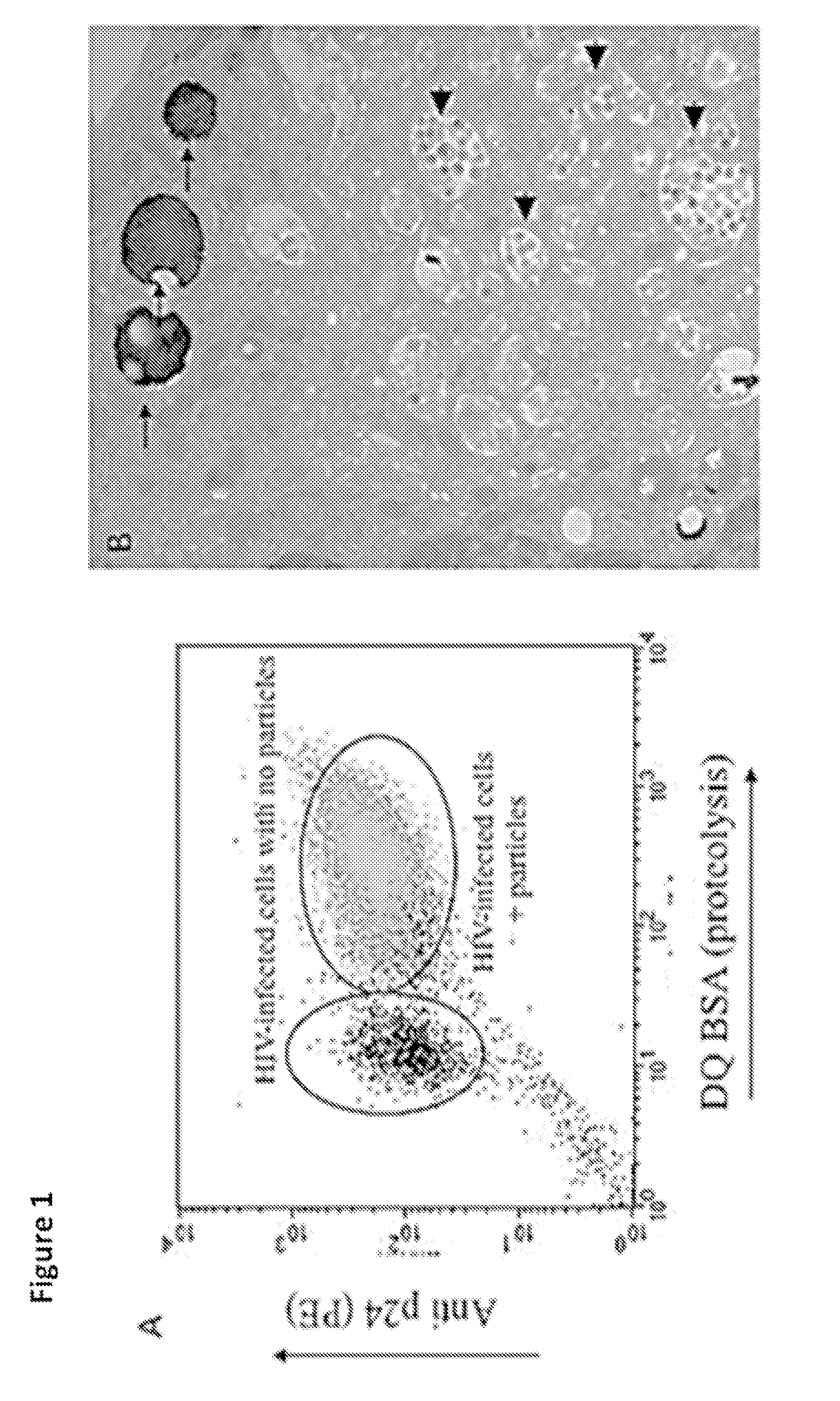
[0043]As shown in FIG. 1, in in vitro infections of human monocyte-derived macrophages with HIV (BaL strain), the infection was easily detected by FISH and flow cyometry staining with anti-p24 antibody. The level of the infection was also visualized by electron microscopy. However, when human alveolar macrophages from HIV positive (HIV+ve) patients were probed with anti-p24 antibodies, the level of viral antigen was below the level of detection by flow cytometry. To enhance the signal a cocktail of antibodies against p24, p17, p120 and Gag was used. But again there was no detectable HIV in cells from HIV+ve patients. These results demonstrated that the viral burden in vivo is considerably lower than that found in vitro.

example 2

[0044]Because of the lack of positive results described in Example 1, we revised our approach to investigate FISH as a technique to improve sensitivity of HIV detection. We engaged in this approach because of published reports that describe using this method successfully to detect HIV in lymphocytes from HIV-infected patients. We first attempted to use a commercially available flow cytometry based in situ HIV-1 mRNA test for intact human cells. The test is offered commercially with the characterization that it can be used in any single cell suspension appropriate for flow cytometry or image analysis, and that it is suitable for use with cell lines, peripheral blood, dissociated tissues, and bodily fluids. It also purports to be able to detect the presence of persistent HIV-1 replication in individuals with undetectable plasma viral load. However, our testing with this commercially available test failed to detect virus in either patients' cells or in 8Ef cells infected with a pseudov...

example 3

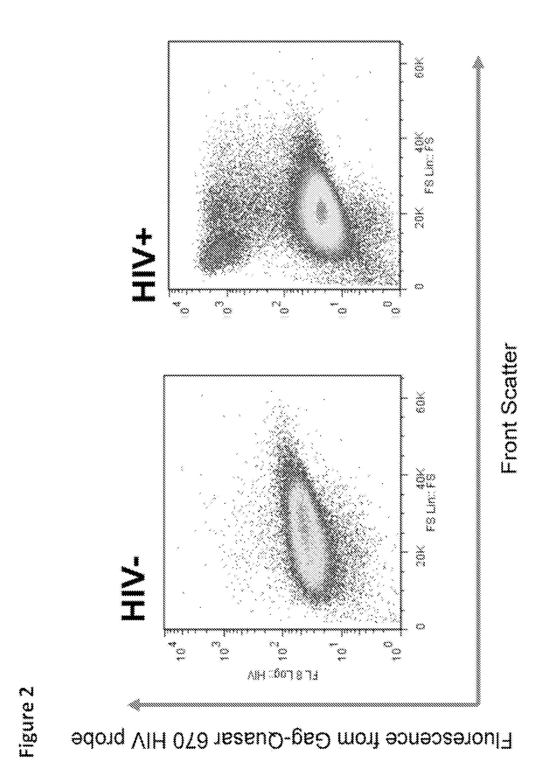
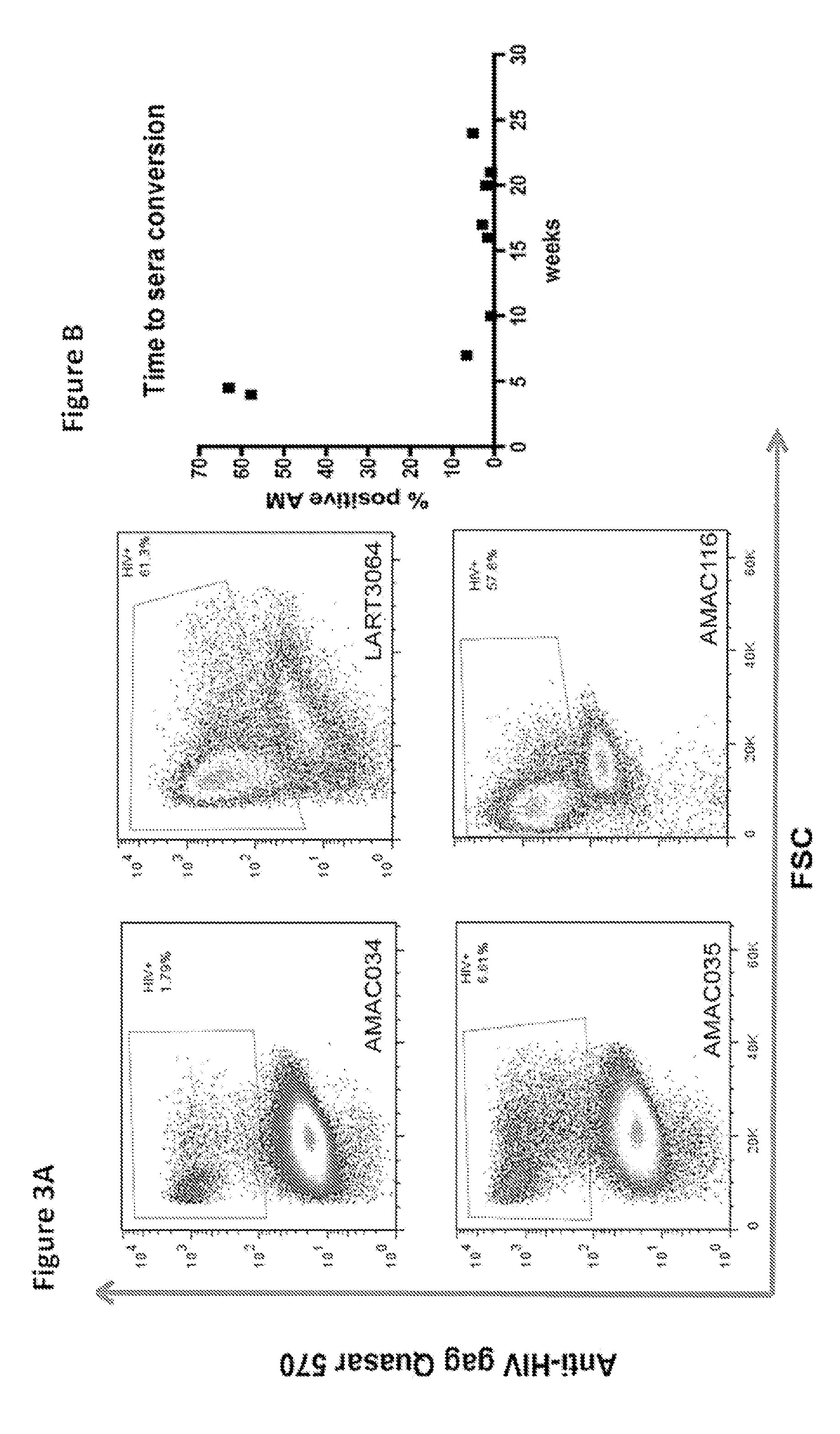
[0045]In order to continue attempting to develop a test with previously unavailable sensitivity we utilized two different sets of fluorescently labeled probes having the sequences presented in Table 1 and Table 2, which we had prepared by Biosearch Technologies, Inc., (Novato, Calif.). We successfully probed for the presence of HIV in human alveolar macrophages from HIV+ve patient samples using the labeled probes using flow cytometry.


PUM

PropertyMeasurementUnit
timeaaaaaaaaaa
timeaaaaaaaaaa
fluorescence in situaaaaaaaaaa

Abstract

Provided is a method for identifying human immunodeficiency virus (HIV) in a sample. The invention involves use of a plurality of fluorescently labeled oligonucleotide probes and flow cytometry to detect the presence or absence of HIV in macrophages that are obtained from a mucosal surface. The invention is particularly useful for detecting HIV infection prior to seroconversion.

Description

CROSS REFERENCE TO RELATED APPLICATIONS[0001]This application claims priority to U.S. Provisional application No. 61 / 563,216, filed on Nov. 23, 2011, the disclosure of which is incorporated herein by reference.STATEMENT REGARDING FEDERALLY SPONSORED RESEARCH[0002]This invention was made with government support under contract no. HL100928 awarded by the National Institutes of Health. The government has certain rights in the invention.FIELD OF THE INVENTION[0003]The present invention relates generally to the detection and diagnosis of human immunodeficiency virus infection, and particularly to detection of human immunodeficiency (HIV) infection during early stages of the acute phase.BACKGROUND OF THE INVENTION[0004]Acute HIV (AHI) infection is the stage of infection prior to seroconversion and proper diagnosis of HIV infection at this stage has remained unresolved with serious implications to public health—AHI is associated with the highest rates of secondary HIV transmission over the...

Claims


Application Information

Patent Timeline
Patent Type & Authority Applications(United States)
IPC IPC(8): C12Q1/70
CPCC12Q1/703C12Q1/70C12Q2543/10
Inventor RUSSELL, DAVID G.MWANDUMBA, HENRYJAMBO, KONDWANI
Owner CORNELL UNIVERSITY
Why Eureka
Try Eureka