
Computational methods and apparatus for meibography

a meibography and computational method technology, applied in the field of meibography, can solve the problems of irregular imaged glands, inconvenient local thresholding and conventional methods such as local thresholding

Inactive Publication Date: 2014-12-11
AGENCY FOR SCI TECH & RES +1
View PDF0 Cites 26 Cited by
  • Summary
  • Abstract
  • Description
  • Claims
  • Application Information

AI Technical Summary

Benefits of technology

The present invention is about automating the processing of images of the eye's ocular region, which contains many meibomian glands. The invention can help identify the location of these glands and get numerical data that describes them. This data can then be used to grade the glands.

Problems solved by technology

On the other hand, for an unhealthy eye, the imaged glands show irregularities.
The IR images have several features that make it challenging to automatically detect the gland regions:1. low-contrast between gland and non-gland regions;2. specular reflections caused from smooth and wet surfaces;3. inhomogeneous gray-level distributions over regions because of thermal imaging;4. irregularities in imaged regions of the ocular surface.
However, because of the above mentioned imaging conditions, conventional methods such as local thresholding are not suitable for partitioning image into gland and non-gland regions.

Method used


Image

  • Computational methods and apparatus for meibography
  • Computational methods and apparatus for meibography
  • Computational methods and apparatus for meibography

Examples


1st embodiment

1.1 GABOR FUNCTIONS

[0036]The first embodiment of the invention is a method of detecting meibomian glands, making use of the family of two-dimensional (2D) Gabor functions. It is known to use a Gabor function as a receptive field function of a cell, to model the spatial summation properties of simple cells [1]. A modified parametrization of Gabor functions is used to take into account restrictions found in the experimental data [2, 3]. Suppose there is a light impulse at a point (x, y) on a 2-dimensional visual field Ω (that is (x, y)∈Ω⊃R2). The Gabor function is denoted by Gλ,θ,ψ(x, y) which is a real valued number (i.e. Gλ,θ,ψ(x, y)∈R). The Gabor function is given by [2]:

Gλ,θ,ψ(x,y)=exp(-x~2+γ2y~22σ2)cos(2πx~λ+ψ)-Gλ,θ,ψDC(1)

where

{hacek over (x)}=(x−x0)cos(θ−π / / 2)÷(y−y0)sin(θ−π / 2),

{tilde over (y)}=−(x−x0)sin(θ−π / 2)+(y−y0)cos(θ−π / 2),

and Gλ,θ,ψDC is DC term due to cosine function. The DC term

Gλ,θ,ψDC=∫∫ΩGλ,θ,ψ(x,y)xy(1)

is subtracted from Gλ,θ,ψ to remove the bias.

[0037]Without loss of...

2nd embodiment

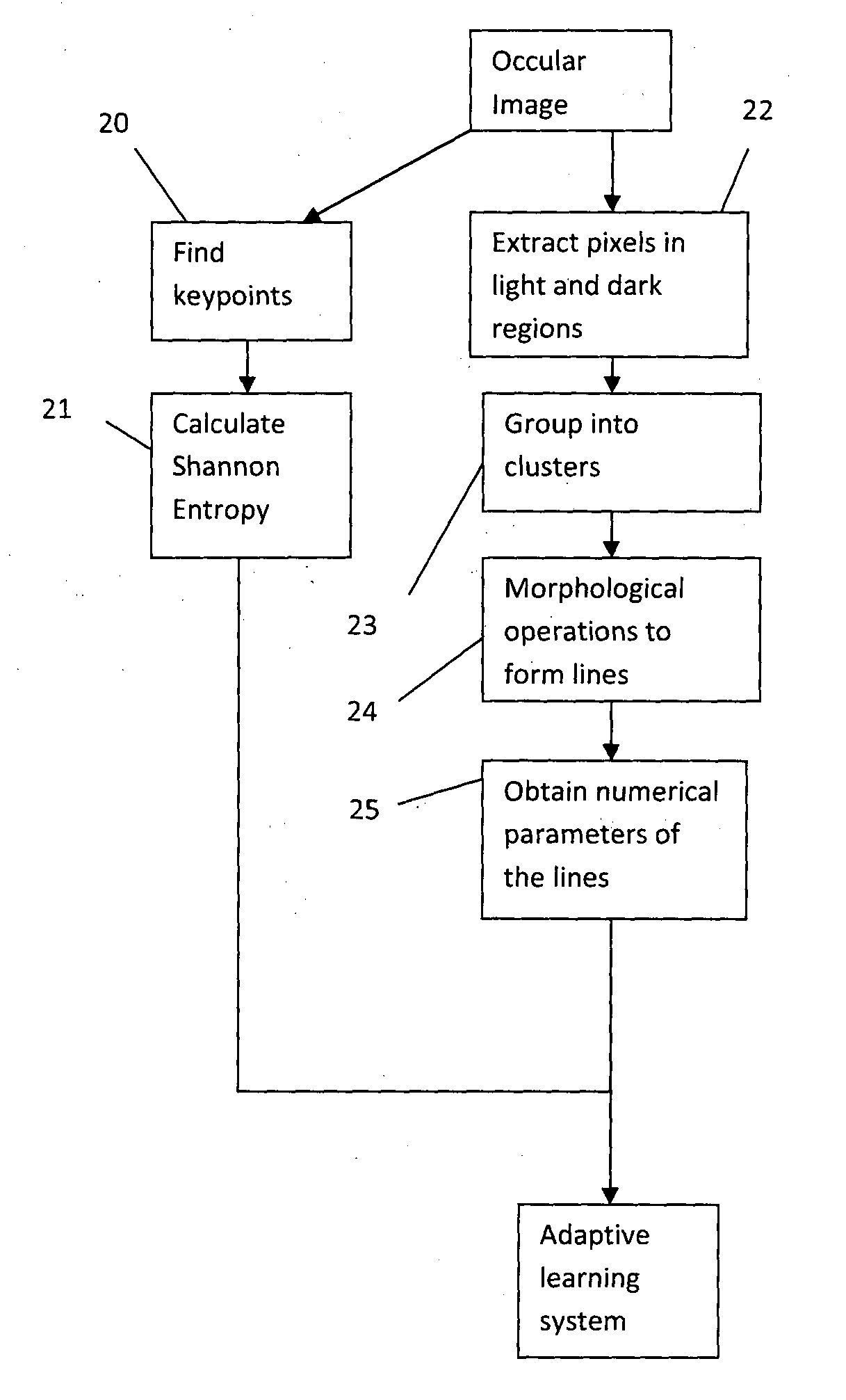
[0054]The second embodiment aims to provide a way of grading a subject, i.e. alloting him into one of at least two categories, such as “healthy”, “unhealthy” or “intermediate”.

[0055]The overall method of the second embodiment is illustrated in FIG. 18. A single occular image is used to obtain one or more numerical parameters (“features”) indicative of whether the image is healthy or not. Note that not all the numerical parameters described below may be collected in realisations of the embodiment, but preferably more than one parameter is collected, and in this case the numerical parameters are combined by an adaptive learning system (such as a support vector machine (SVM) which has been subject to supervised learning), to generate an output indicative of whether the image is healthy or not.


PUM


Abstract

An occular image of a region including meibomian glands is processed automatically. The processing may derive a grade indicative of the health of the meibomian glands, by using in the occular image to obtain one or more numerical parameters characterizing the meibomian glands shown in the occular image, and determining the grade using the one or more numerical parameters. The numerical parameters include a parameter characterizing the diversity between scale parameters of significant features of the image obtained by a scale-space transform, and / or parameters obtained by measurement of lines in the occular image representing respective glands. Meibomian glands can be identified on ocular images using Gabor filtering as a local filtering technique. The parametrization in shape, local spatial support, and orientation of Gabor filtering is particularly suitable for detecting meibomian glands.

Description

FIELD OF THE INVENTION[0001]The present invention relates to computational methods and apparatus for processing images of the meibomian glands to derive information characterizing abnormalities in the glands, indicative of medical conditions.BACKGROUND OF THE INVENTION[0002]The meibomian glands are sebaceous glands at the rim of the eyelids inside the tarsal plate, responsible for the supply of meibum, an oily substance that prevents evaporation of the eye's tear film. Meibum is a lipid which prevents tear spillage onto the cheek, trapping tears between the oiled edge and the eyeball, and makes the closed lids airtight. It further covers the tear surface, and prevents water in the tears from evaporating too quickly. Dysfunctional meibomian glands can cause dry eyes (since without this lipid, water in the eye evaporates too quickly) or blepharitis; and other medical conditions.[0003]It is known to capture infra-red (IR) images of ocular surface to analyse the morphological structures...

Claims


Application Information

Patent Timeline
Patent Type & Authority Applications(United States)
IPC IPC(8): G06T7/00G06K9/62G06K9/46
CPCG06T7/0012G06T2207/30041G06K9/62G06K9/46G06V40/197G06V40/193G06V10/449G06V10/462
Inventor LEE, HWEE KUANKOH, PATRICKCELIK, TURGAYTONG, LOUIS HAK TIENPETZNICK, ANDREA
Owner AGENCY FOR SCI TECH & RES
Learn More