Method For Processing Blood Sample

a blood sample and processing method technology, applied in the field of blood sample processing, can solve the problems of difficult detection or obtaining cells found in blood at much lower levels, limited ability of cellsearch system to capture cancer cells, and inability to capture cells by cellsearch system, so as to reduce the filtration area and improve the capture rate of rare cells

Active Publication Date: 2015-03-26
ARKRAY INC
View PDF2 Cites 6 Cited by
  • Summary
  • Abstract
  • Description
  • Claims
  • Application Information

AI Technical Summary

Benefits of technology

[0026]Non-Patent Documents 6 to 8 disclose a method for separating CTCs in view of the fact that (i) the cancer cells are generally larger than the blood cells and (ii) the inherent flexibility and deformability of the blood cells allow the white blood cells to pass through holes smaller than themselves. However, the method uses a filter with non-uniform holes, and some of them are fused together into large holes. Thus, the cancer cells can go through these holes, which leads to a low capture rate.
[0050]According to the present disclosure, in one or more embodiments, not only deformable cancer cells that would easily pass through the filter, but also small cancer cells can be captured alive while reducing the filtration area. Moreover, according to the present disclosure, in one or more embodiments, the capture rate of rare cells can be improved when the rare cells are present in the blood sample.

Problems solved by technology

Conventionally, with respect to blood and CTCs, the predominance of red blood cells and white blood cells has made it difficult to detect or obtain cells that are found in blood at much lower levels.
Therefore, the ability of the CELLSEARCH system to capture cancer cells is limited, since it utilizes affinity and relies on the amount of antigen expressed.
However, due to the reliance on the amount of surface antigen expressed by CTCs, some cells cannot be captured by the CELLSEARCH system.
Thus, even if CTCs are actually traveling in blood, the CELLSEARCH system may not capture many of them and results in low sensitivity.
However, the method uses a filter with non-uniform holes, and some of them are fused together into large holes.
Thus, the cancer cells can go through these holes, which leads to a low capture rate.

Method used


Image

  • Method For Processing Blood Sample
  • Method For Processing Blood Sample
  • Method For Processing Blood Sample

Examples


example 1

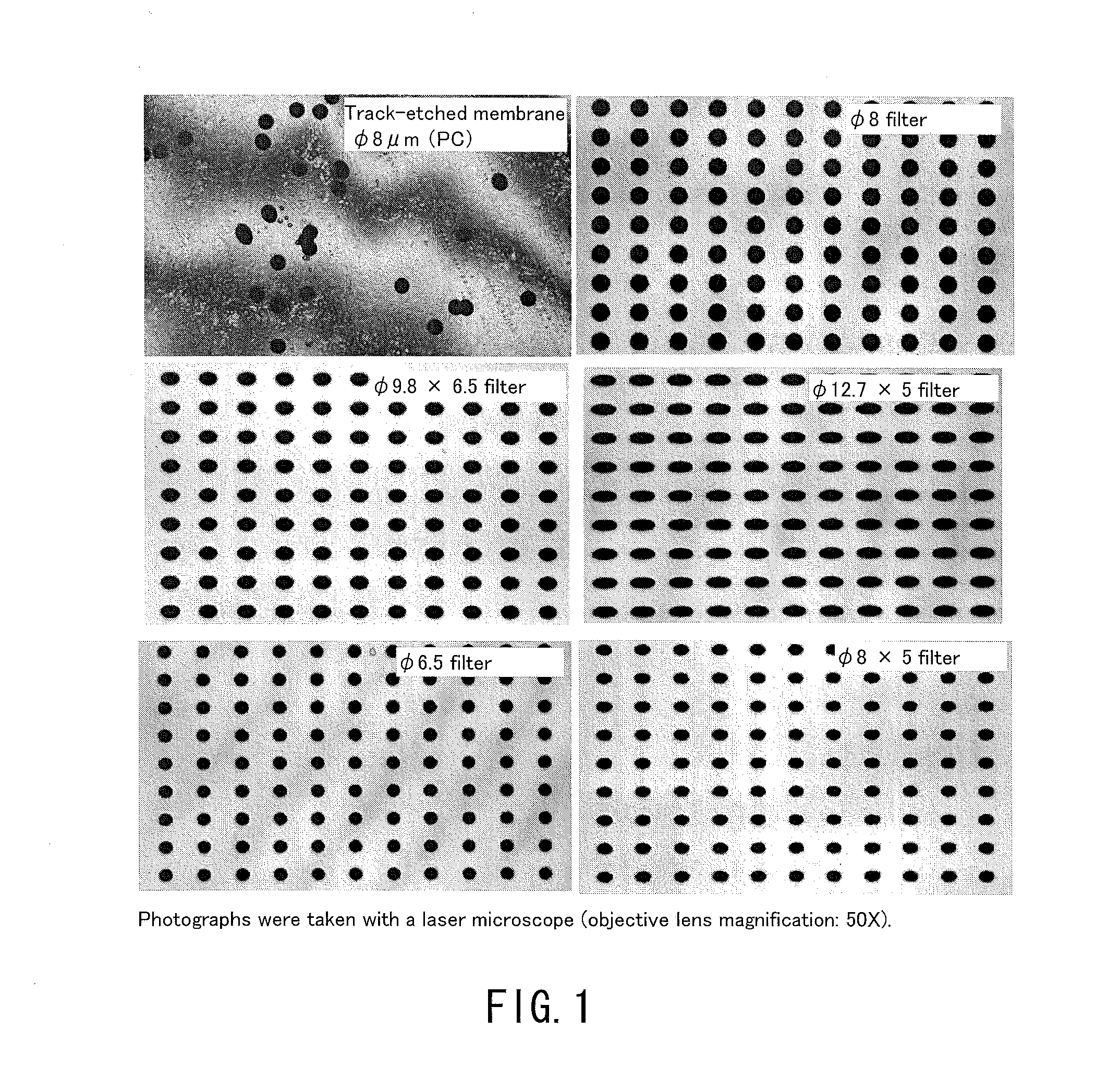
[0141]In the following examples, using filters having the same number of holes and the same filtration area, blood samples were processed in a volume of 14 nl per hole (the total volume of the blood sample to be processed: 1 ml). Consequently, the recovery rate of small cells that would pass through 10 μm holes was generally high as long as the holes were smaller than the cells regardless of the hole shape. On the other hand, the recovery rate of deformable cancer cells was low when the holes were small. Table 3 and FIG. 3 show data comparing the capture capabilities of various holes among the filters. In this case, the total volume of the blood sample to be processed was 1 ml. As shown in Table 3, whether the hole shape was a true circle or an ellipse, the capture rate of deformable cancer cells was significantly reduced as the hole area became smaller. Moreover, clogging (blockage) occurred in φ 4 μm holes before the blood sample in a volume of 14 nl / hole had reached the total vol...

example 2

[0142]The amount of a blood sample to be processed through each of the filters was considered. If a large amount of blood can be processed with the same filtration area, it is possible to reduce the filtration area of the filter to be used, to suppress the bulk of the filtration portion in the device, and to ensure cost effectiveness, as compared to the conventional examples. Tables 4 and 5 show average data of the capture rates of SW620 cells and SNU-1 cells, respectively. The average data are also plotted in FIG. 4. In this case, the φ 8 filter, the φ 9.8×6.5 filter, and the φ 12.7×5 filter were used, each of which achieved a better capture rate for 1 ml of blood, and the amount of the blood sample to be processed was increased up to 25 ml. Surprisingly, when the filtration area was 20 mm2, these filters continued to process blood without clogging (blockage) until 25 ml (filtration time: 50 min to 70 min). Thus, a high capture rate of small cancer cells was maintained (see Table 4...

example 3

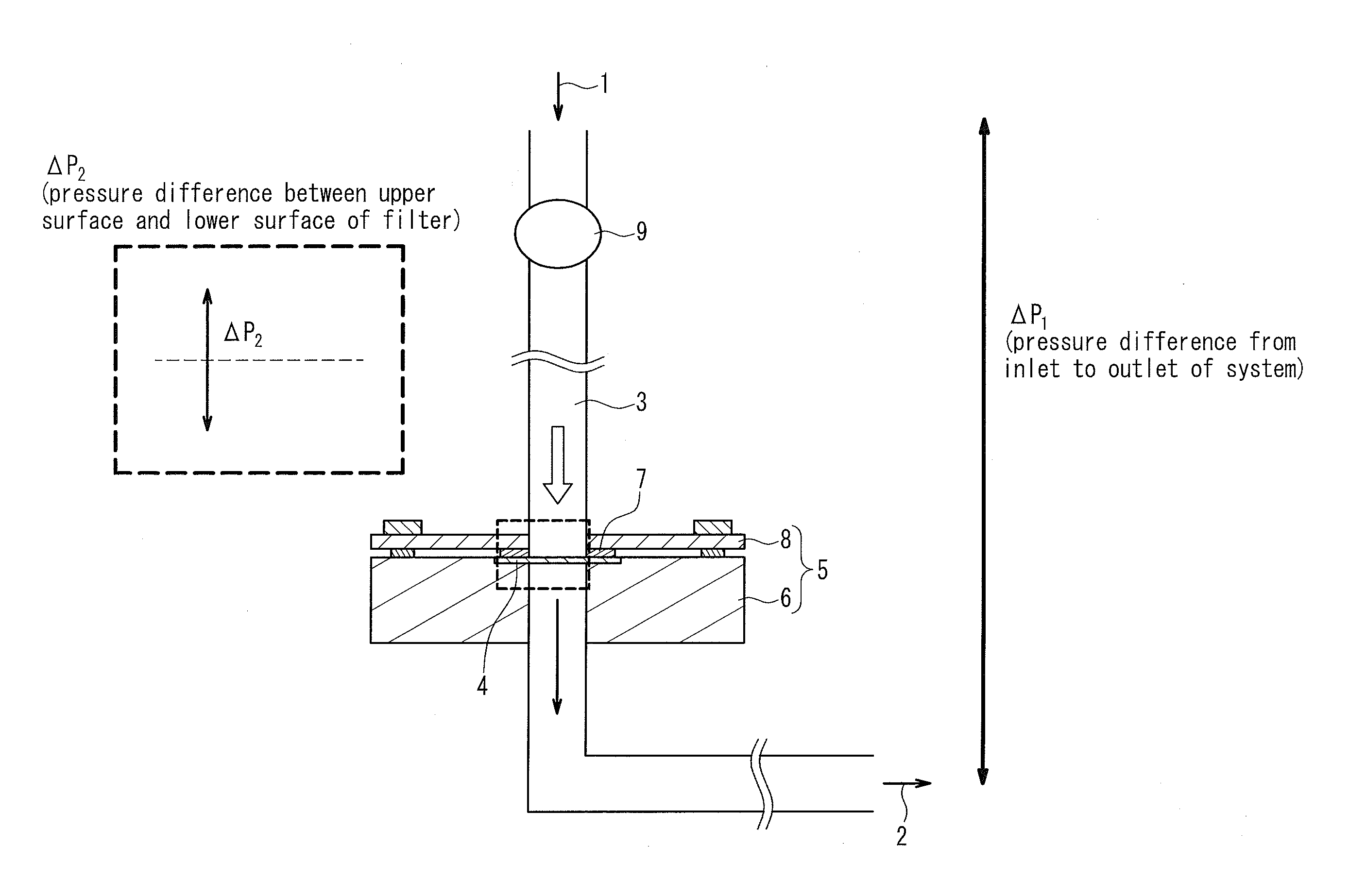
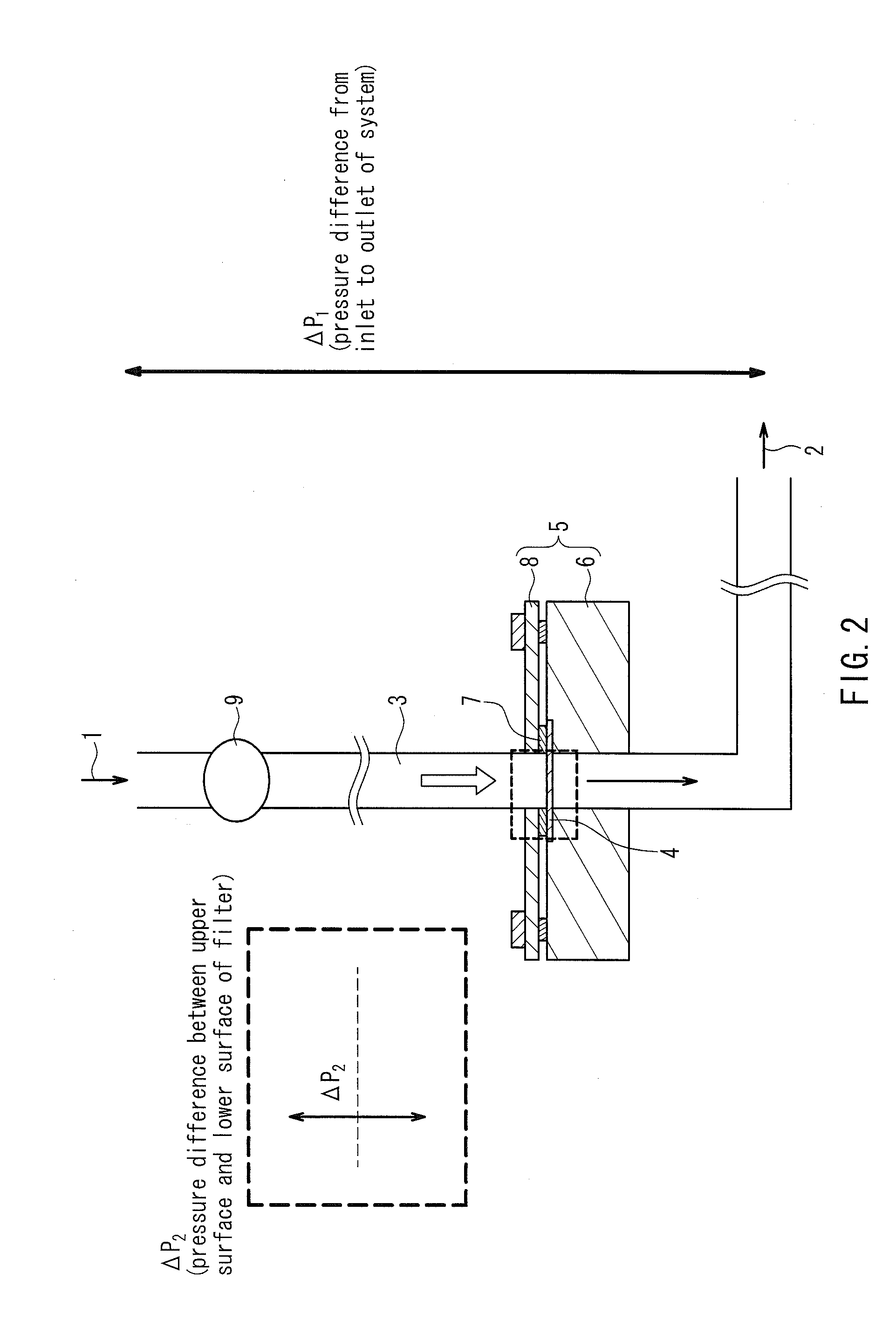
[0144]Next, data of the capture rates of cells when the filtration pressure was varied during filtration will be shown in the following. In general, high pressure would make it easy for cells to pass through the holes of a filter, and thus degrade the capture capabilities, and low pressure would make it difficult for cells to pass through the holes of a filter, and thus be expected to improve the capture rate. However, as shown in Table 7-1, when the amount of blood per hole was 108 nl, the capture rate was not stable and reduced particularly on the lower pressure side of the φ 8 filter and the φ 9.8×6.5 filter. It is considered that since the filtration time was too long, the cells passed through the filters due to their viscoelasticity.

[0145]Next, data obtained by varying the filtration pressure with respect to 108 nl of a blood sample to be processed per hole (the total volume of the blood sample to be processed: 8 ml) and 14 nl of a blood sample to be processed per hole (the tot...


PUM

PropertyMeasurementUnit
diameteraaaaaaaaaa
volumeaaaaaaaaaa
volumeaaaaaaaaaa

Abstract

A method for processing a blood sample is provided that can improve the recovery rate of deformable rare cells that would easily pass through a filter and small rare cells while reducing the filtration area of the filter, and that can recover the rare cells alive.

Description

BACKGROUND OF THE INVENTION[0001]1. Field of the Invention[0002]The present disclosure relates to a method for processing a blood sample, a method for separating rare cells in a blood sample, a method for enumerating or analyzing circulating tumor cells in blood, a filter used in these methods, and a device for capturing rare cells.[0003]2. Description of Related Art[0004]The blood cell components are mainly red blood cells, white blood cells, and platelets. In some cases, cells other than these blood cells may be found in blood. Circulating tumor cells (CTCs) are one example of such cells. Metastasis of cancer is attributed to the transfer and growth of cancer cells to a distant site in the body through the blood or lymphatic vessels. It has been reported that there is a relationship between the number of CTCs in blood and the risk and prognosis of metastasis of cancer. The enumeration of CTCs in blood and the measurement of the nucleic acids of CTCs are known to be used as indicat...

Claims


Application Information

Patent Timeline
Patent Type & Authority Applications(United States)
IPC IPC(8): G01N33/50B01D29/03B01D35/30
CPCG01N33/5094B01D29/03B01D35/30B01D63/087G01N33/491
Inventor TAKAGI, HIDENORI
Owner ARKRAY INC
Why Eureka
Try Eureka