
Patient-specific analysis of positron emission tomography data

a tomography and patient technology, applied in the field of medical imaging, can solve problems such as poor reproducibility

Active Publication Date: 2015-03-26
BLACK RICHARD R
View PDF15 Cites 10 Cited by
  • Summary
  • Abstract
  • Description
  • Claims
  • Application Information

AI Technical Summary

Benefits of technology

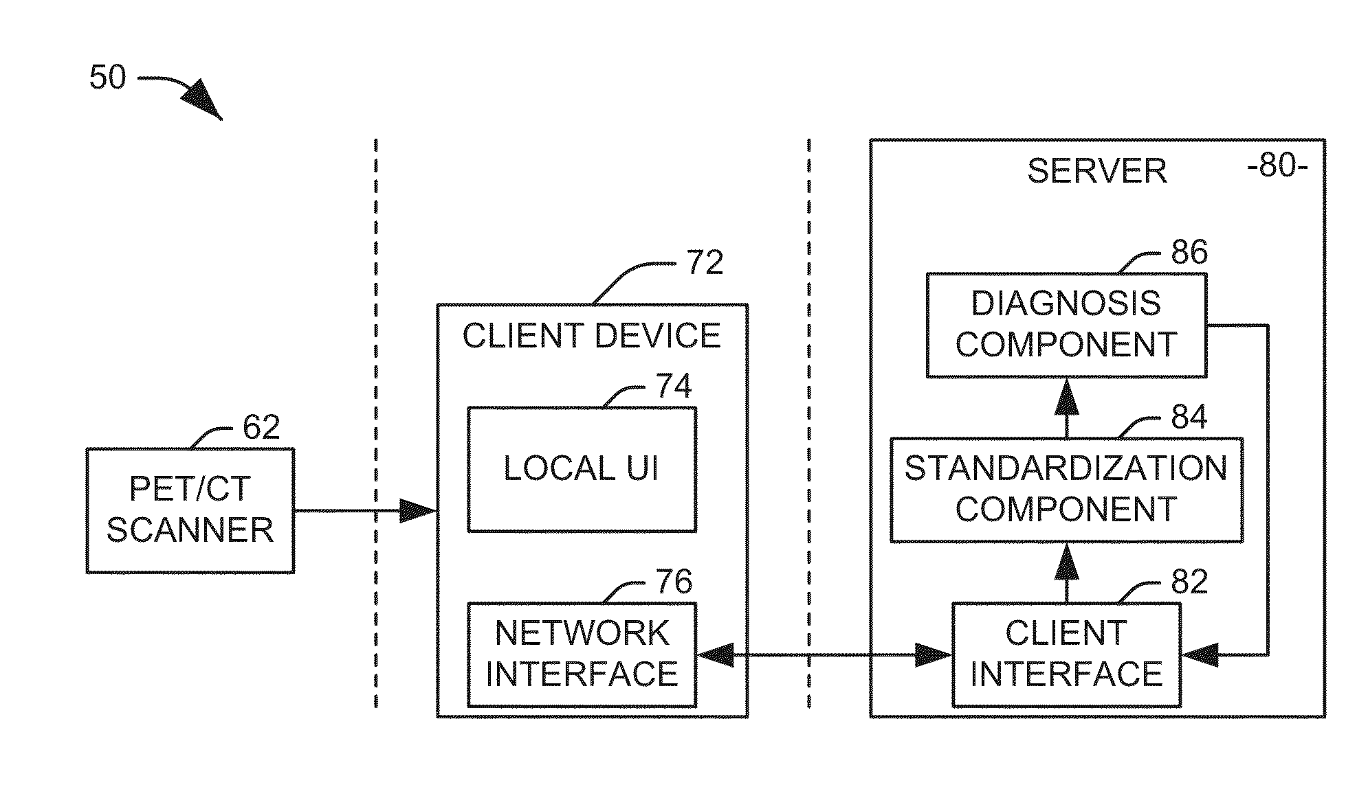
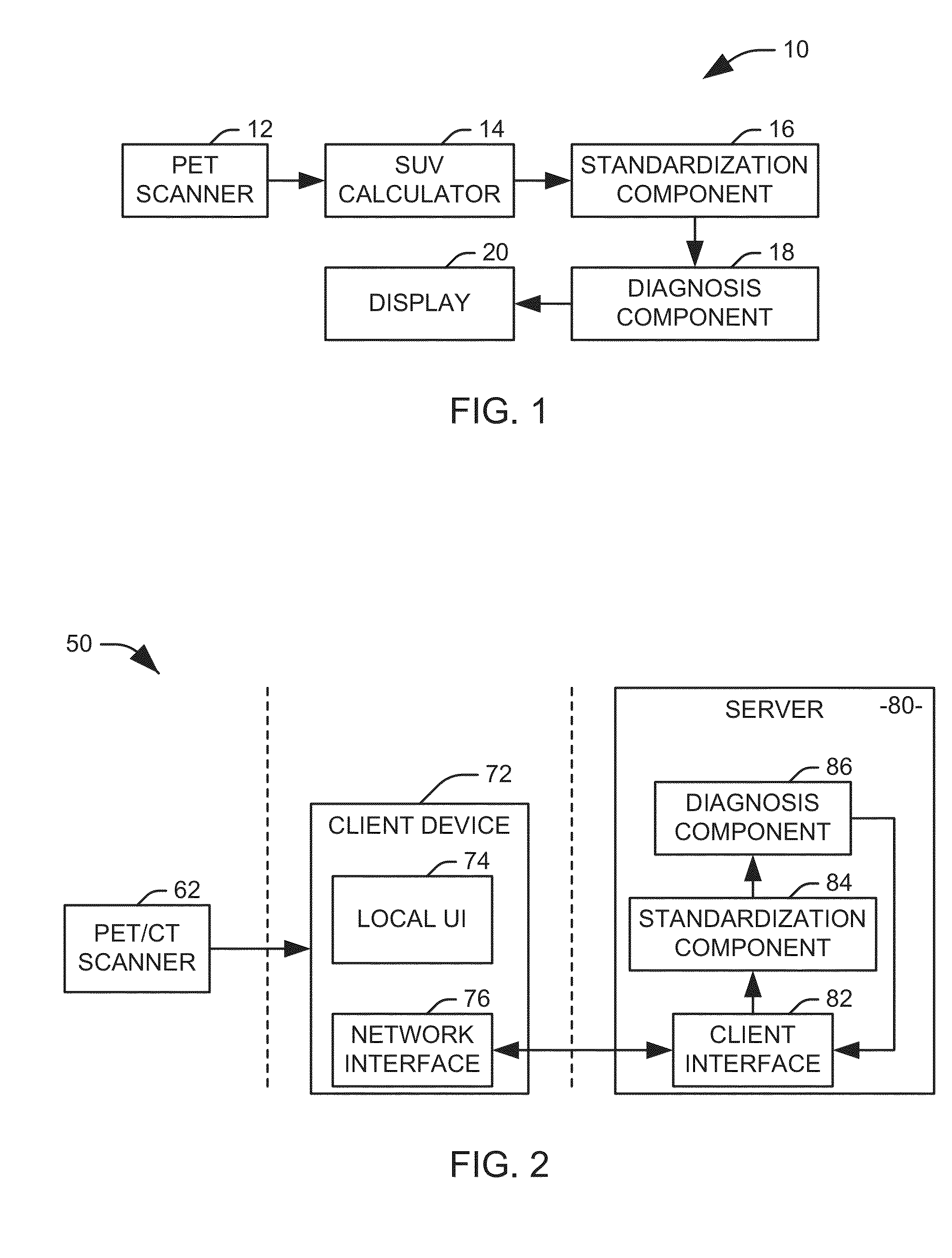
This patent describes a system and method for evaluating abnormal glucose uptake in tissue within a patient. The system includes a positron emission tomography (PET) scanner to obtain a set of PET images of the tissue of interest and the liver. A standard uptake value (SUV) calculator is used to calculate a set of SUVs for the tissue and liver. A standardization component applies a correction value to the set of SUVs based on information from the liver. A diagnosis component compares the set of SUVs to a decision threshold to determine if the glucose uptake in the tissue of interest is abnormal. The detection of abnormal glucose uptake can aid in the diagnosis and treatment of various medical conditions.

Problems solved by technology

Such a qualitative approach is subjective and may be associated with poor reproducibility.

Method used


Image

  • Patient-specific analysis of positron emission tomography data
  • Patient-specific analysis of positron emission tomography data
  • Patient-specific analysis of positron emission tomography data

Examples


Embodiment Construction

[0016]One widely used SUV threshold for the categorization of neoplastic versus non-neoplastic disease is considered to be 2.5. It has been suggested, however, that different thresholds for the designation of malignant disease based on the location of involvement, so that the commonly utilized value of 2.5 may be associated with an overall decrease in specificity. To further complicate the use of the SUV in the evaluation of individuals with suspected malignant disease, variability in the SUVs between and within individuals may occur on the same imaging device. The use of serial examinations on the same patient using different devices further increases the inconsistency and reduces the overall accuracy of the individual examination.

[0017]In accordance with an aspect of the present invention, systems and methods are provided for patient-specific analysis of positron emission tomography (PET) images. Specifically, the inventor has determined that the stability of glucose uptake in the...


PUM


Abstract

Systems and methods are provided for evaluating tissue for abnormal glucose uptake within a region of interest within a patient. A PET scanner obtains PET imagery including the region of interest and at least part of the liver. An SUV calculator calculates a first set of SUVs representing the region of interest and a second set SUVs representing the liver from the set of at least one PET image. A standardization component calculates a correction value as a function of the second set of SUVs and applies the correction value to either a decision threshold associated with the region of interest or the first set of SUVs. A diagnosis component compares the first set of SUVs to the decision threshold to determine if the glucose uptake within the region of interest is abnormal. A display provides the determination to a user.

Description

RELATED APPLICATIONS[0001]This application is claims priority to each of U.S. Provisional Patent Application Ser. No. 62 / 054,661 filed Sep. 24, 2014, U.S. Provisional Patent Application Ser. No. 61 / 984,246 filed Apr. 25, 2014, and U.S. Provisional Patent Application Ser. No. 61 / 882,261 filed Sep. 25, 2013. Each of these applications is hereby incorporated by reference.BACKGROUND OF THE INVENTION[0002]1. Field of the Invention[0003]The invention relates generally to medical imaging, More specifically, the invention relates to patient-specific analysis of positron emission tomography data.[0004]2. Background of the Invention[0005]The early diagnosis, appropriate staging, and the subsequent assessment of treatment outcomes of various malignancies can be enhanced by modern imaging techniques. In 1956, Warburg suggested that many malignant cells utilize anaerobic consumption of glucosepyruvate, as the primary energy source. Two decades later, Bustamonte et al proposed using radiolabeled ...

Claims


Application Information

Patent Timeline
Patent Type & Authority Applications(United States)
IPC IPC(8): A61B6/00A61B6/03
CPCA61B6/50A61B6/032A61B6/5217A61B6/037A61B6/5205G16H50/30
Inventor BLACK, RICHARD R.
Owner BLACK RICHARD R
Learn More