Use of stem cells to prevent neuronal dieback
a stem cell and neuronal technology, applied in the direction of biocide, cardiovascular disorder, drug composition, etc., can solve the problems of high concentration of inhibitory myelin breakdown products, high controversy over the role of neuroinflammation in regeneration failure, and increase the size of lesion, so as to reduce neuronal injury, reduce neuronal injury, and reduce the adhesion of ed-1+ cells
- Summary
- Abstract
- Description
- Claims
- Application Information
AI Technical Summary
Benefits of technology
Problems solved by technology
Method used
Image
Examples
example i
[0158]“Glial Scar Model” Aggrecan-laminin opposing spot gradients (Tom et al., 2004; Steinmetz et al., 2005). These references are incorporated by reference for teaching the glial scar model. This model provides an assay for the effectiveness of cells, proteins, medium, etc., in reducing adhesion / retraction in vitro.
[0159]PGs can induce the so-called dystrophic state in axons if the inhibitory matrix is presented in a spatial organization that more closely resembles that which develops after lesions in vivo. To do this, spots of a solution of the PG aggrecan and the growth-promoting molecule laminin were placed on nitrocellulose coverslips and air dried.
[0160]A consistent artifact of drying produced a crude gradient in which the rim of the spot contained an increasingly higher concentration of aggrecan than in the center. The very outermost part of the rim contained a lower concentration of laminin than any more central region, The optimal ECM concentrations (0.7 mg / ml aggrecan and ...
example ii
Axonal Retraction and Microphages
Summary
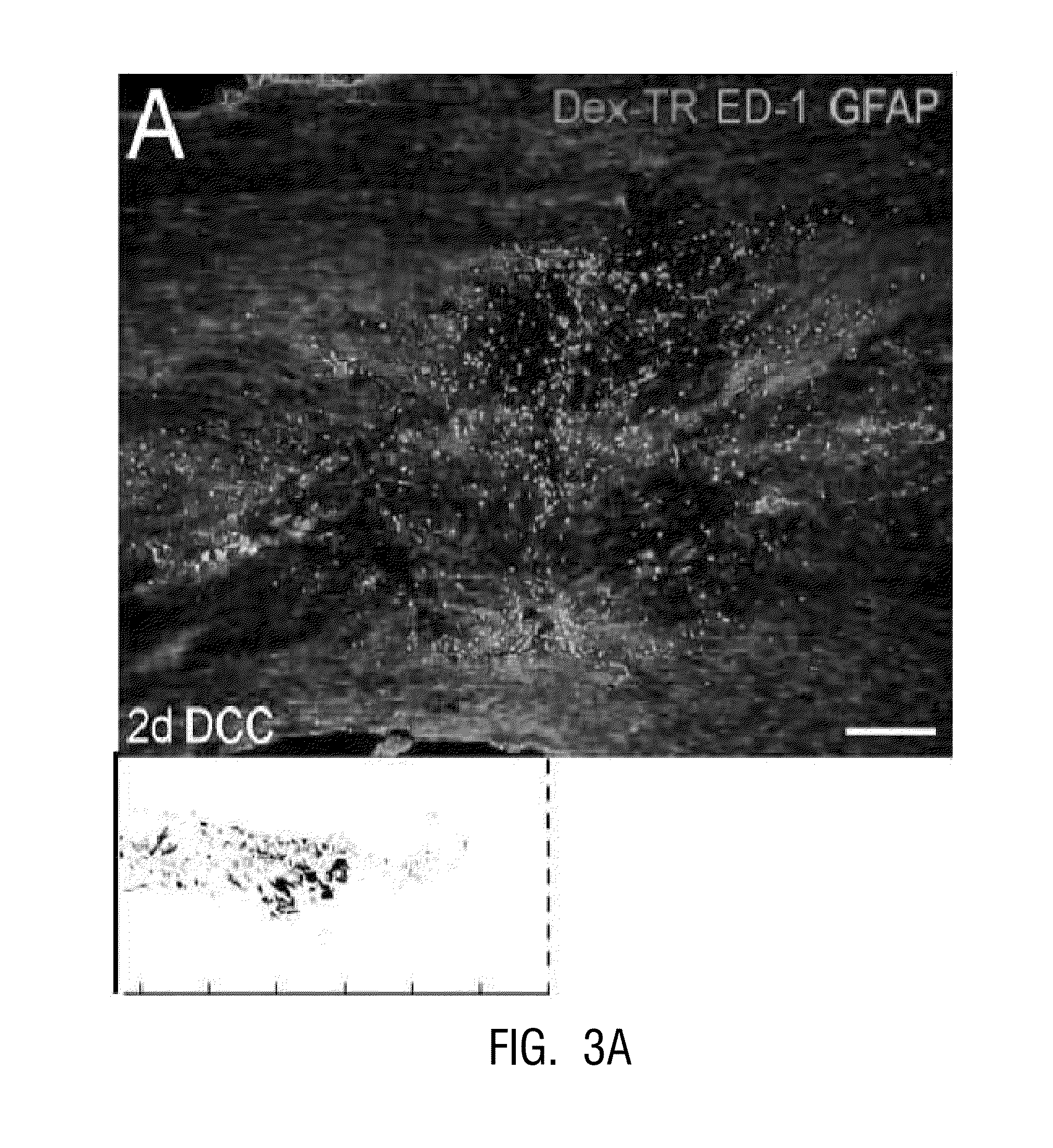
[0161]In vivo, a close correlation was sound between dystrophic retraction clubs at the ends of severed axons and ED-1+ cells following a dorsal column crush spinal cord injury (FIG. 3). The in vitro model of the glial scar (Tom et al., 2004; Steinmetz et al., 2005) was applied to examine the interactions between axons and ED-1+ cells in real time. Direct cell-cell contact between dystrophic growth cones and ED-1+ macrophages induced a long distance axonal retraction (FIGS. 4, 5). The result of using clodronate liposomes (Popovich, et al., 1999) for macrophage depletion in viva was significant reduction in axonal retraction in the clodronate-treated animals compared to controls. These data indicate that ED-1+ cells are directly responsible for retraction of injured spinal cord axons through physical cell-cell interactions.
Results
1. Ascending Dorsal Column Sensory Axons Retract Extensively Following Spinal Cord Injury
[0162]It was considered by ...
example iii
Stem Cells can Prevent Adhesion of Activated Macrophages to DRGs
Results
1. Model
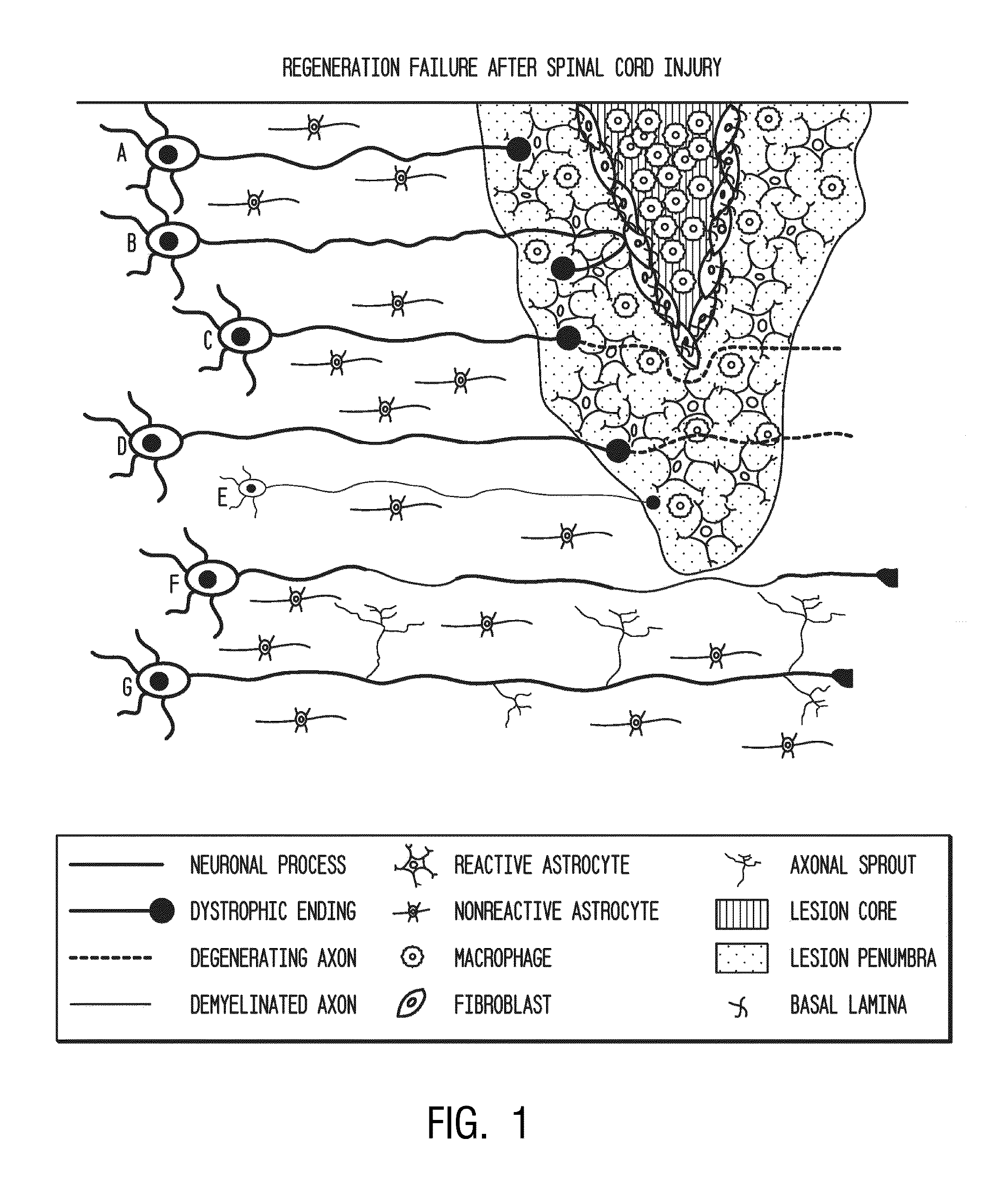
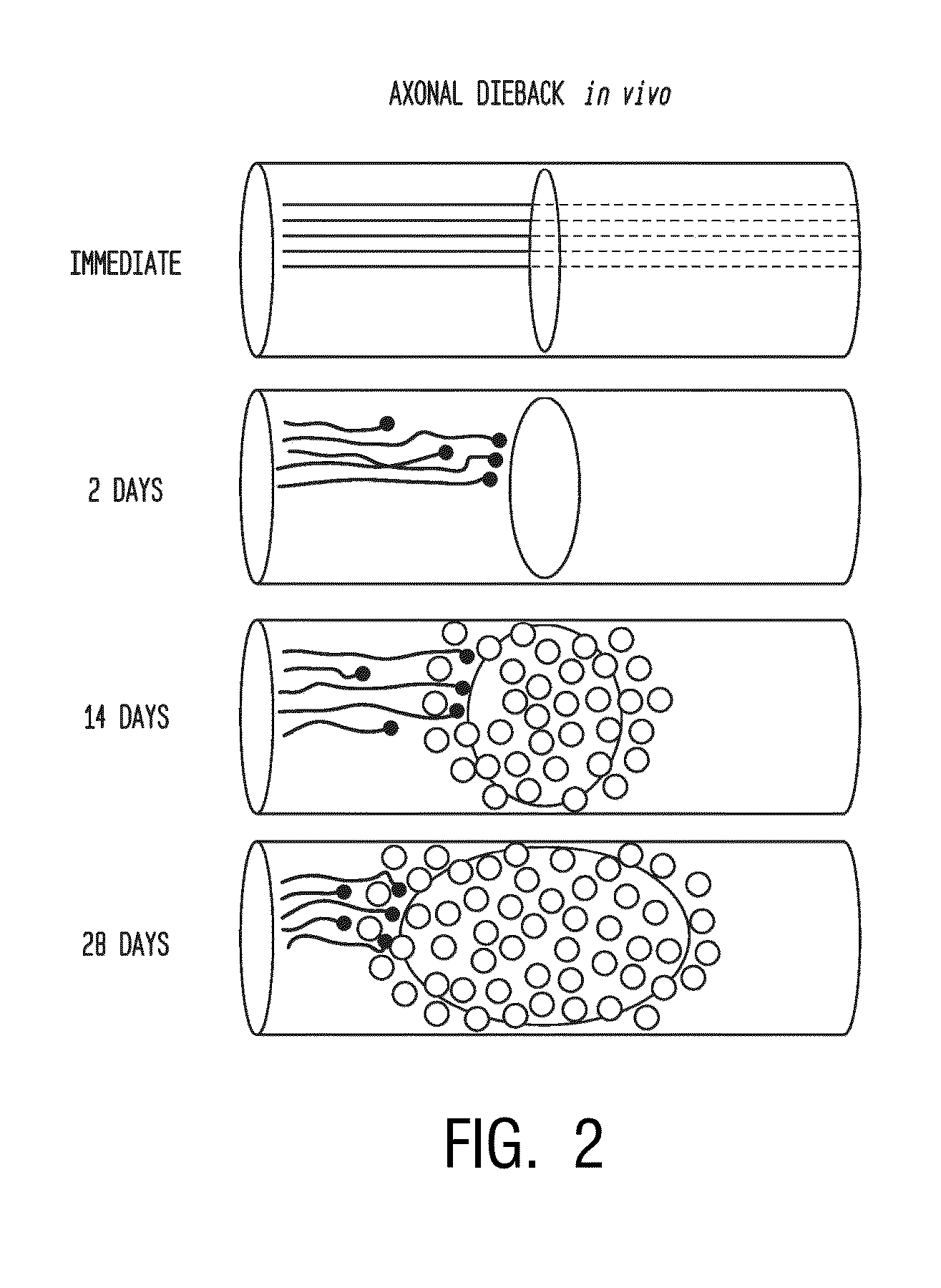
[0191]Following dorsal column crush injury, regenerating axons encounter macrophages and microglia and form dystrophic endings. This is shown schematically in FIG. 1. Previous work from the inventors' laboratory has shown that macrophage infiltration is correlated with axonal dieback following dorsal column crush injury (FIGS. 2-3C). After characterizing the extent of axonal dieback of the ascending dorsal column sensory axons following injury, the inventors established an in vitro model of dieback, which can he used to evaluate various treatment strategies. The in vitro assay consists of cultured adult DRG neurons on a substrate of opposing gradients of the growth-promoting protein laminin and the potently inhibitory chondroitin sulfate proteoglycan aggrecan (Tom et al., 2004). This spot gradient is sufficient to stall axonal growth and induce the formation of dystrophic growth cones like those observed ...
PUM
 Abstract
Description
Claims
Application Information
 - R&D
- Intellectual Property
- Life Sciences
- Materials
- Tech Scout
- Unparalleled Data Quality
- Higher Quality Content
- 60% Fewer Hallucinations
Browse by: Latest US Patents, China's latest patents, Technical Efficacy Thesaurus, Application Domain, Technology Topic, Popular Technical Reports.
© 2025 PatSnap. All rights reserved.Legal|Privacy policy|Modern Slavery Act Transparency Statement|Sitemap|About US| Contact US: help@patsnap.com


