Medical system
- Summary
- Abstract
- Description
- Claims
- Application Information
AI Technical Summary
Benefits of technology
Problems solved by technology
Method used
Image
Examples
first embodiment
[0075]A medical system according to a first embodiment of the present invention will be described below with reference to the drawings.
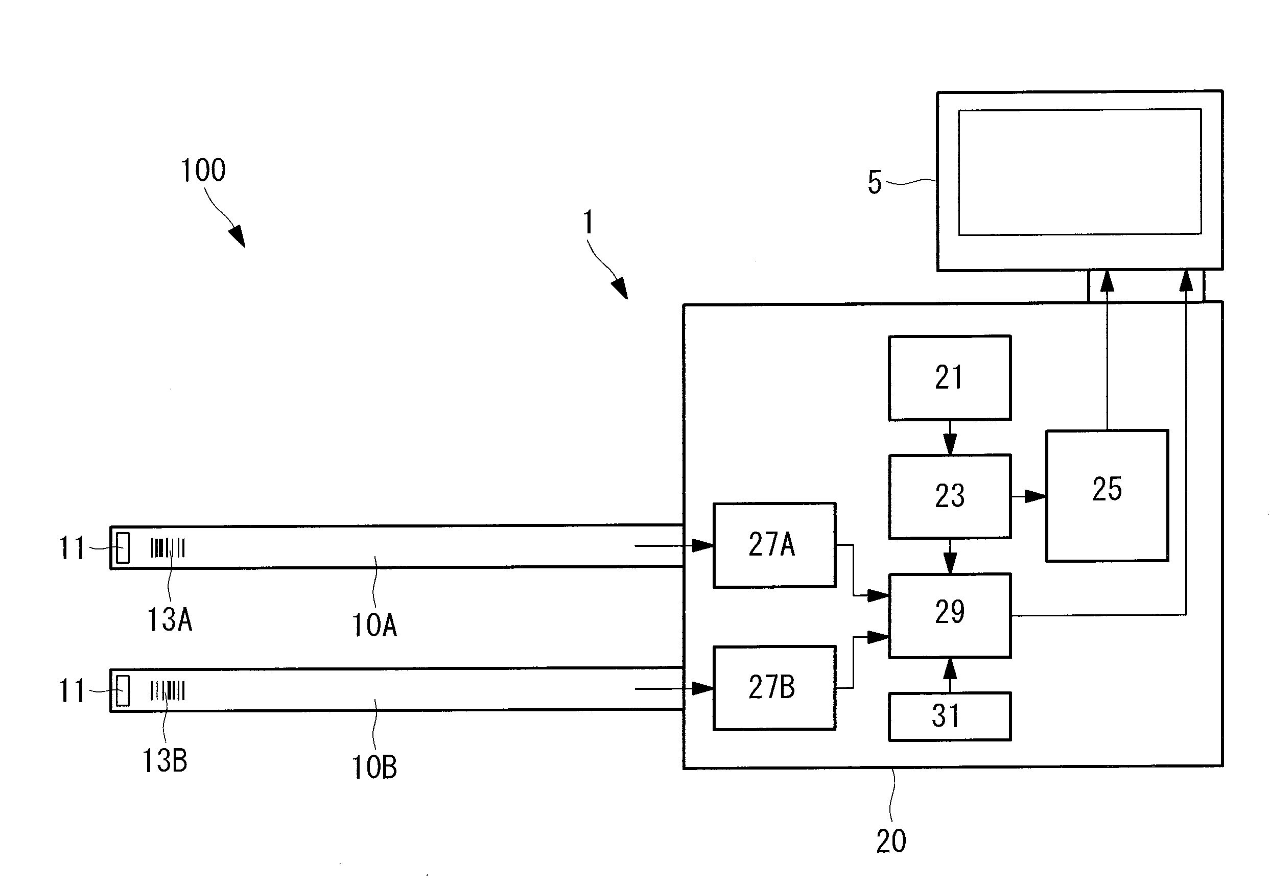
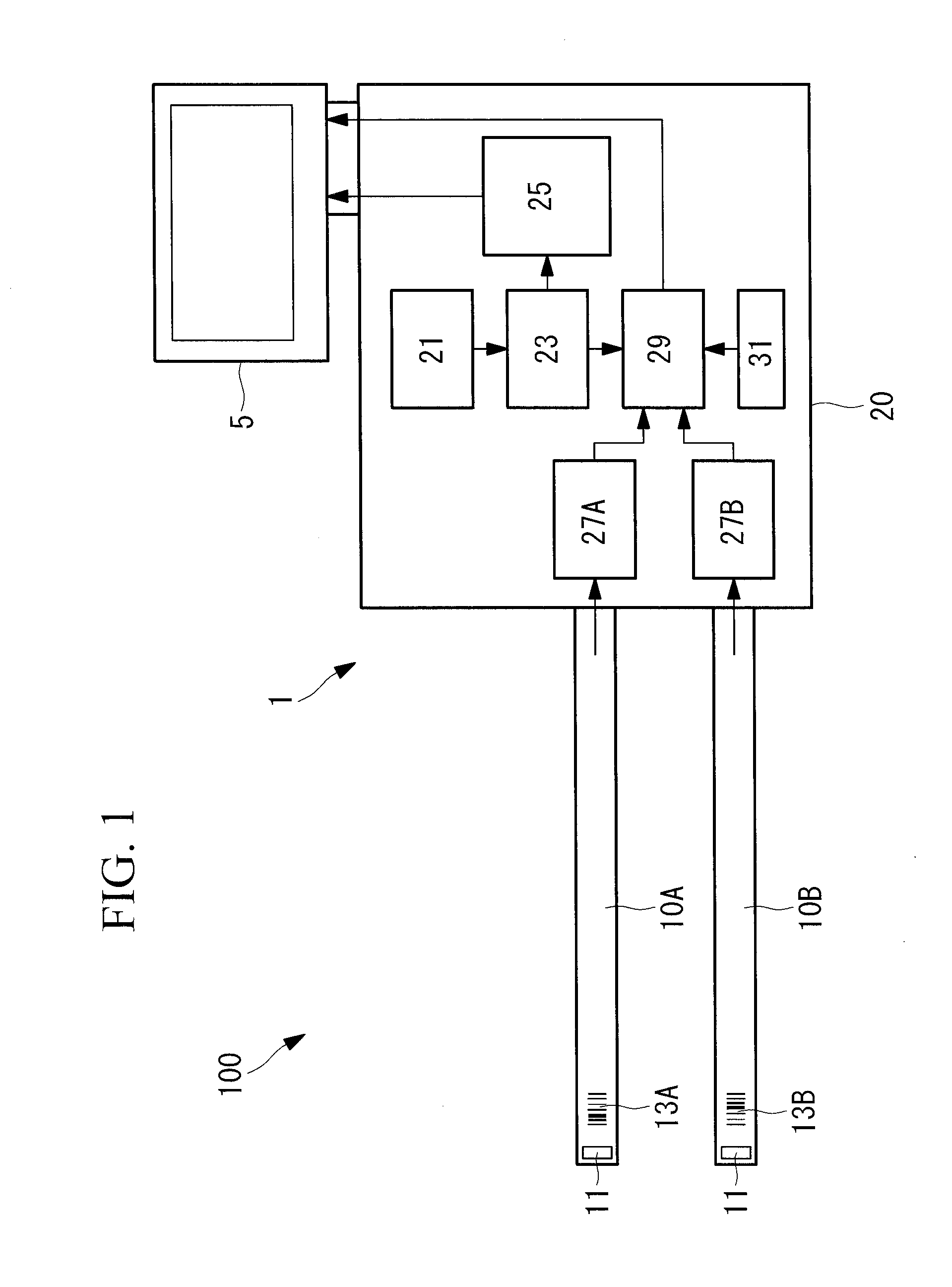
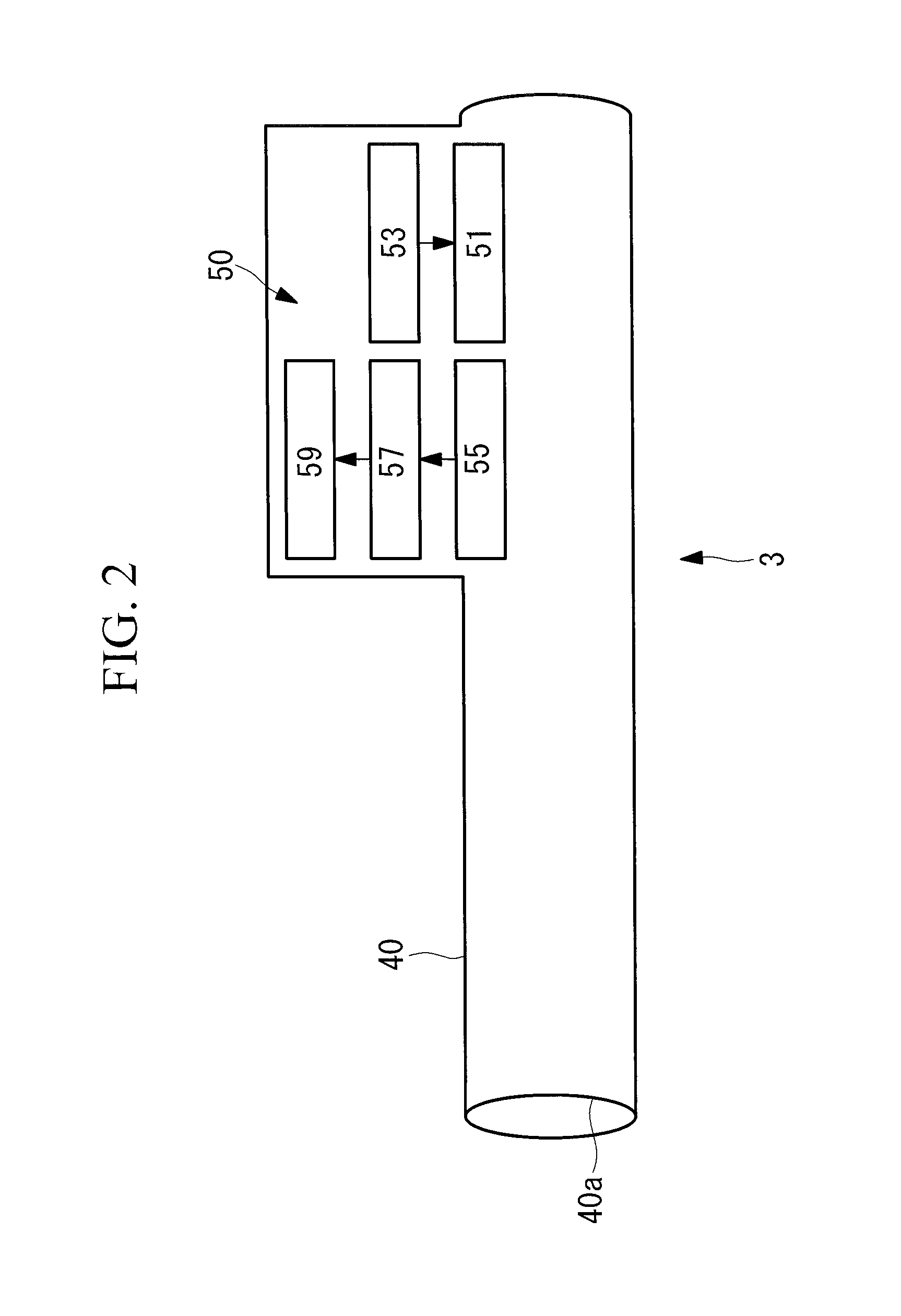
[0076]As illustrated in FIGS. 1 and 2, a medical system 100 according to this embodiment includes an endoscope device (medical device) 1 including a plurality of insertion portions 10A and 10B that are insertable into a body cavity in a biological subject; a sheath unit (outer sleeve) 3 that is attached to the biological subject and guides the insertion portion 10A or 10B of the endoscope device 1 into a body cavity in the biological subject; and a monitor (display unit) 5 that displays images, etc. acquired by the endoscope device 1.
[0077]The endoscope device 1 includes the two endoscope insertion portions 10A and 10B and a main unit 20 that supports the insertion portions 10A and 10B. The insertion portions 10A and 10B are long and substantially cylindrical and have bases that are fixed to or detachable from the main unit 20. The insertion portions...
second embodiment
[0106]A medical system according to a second embodiment of the present invention will now be described.
[0107]As illustrated in FIG. 12, a medical system 200 according to this embodiment differs from the first embodiment in that the insertion portions 10A and 10B are respectively provided with RFID (radio frequency identification) tags 113A and 113B that are capable of outputting unique identification information (insertion-portion identification information) instead of the barcodes 13A and 13B.
[0108]Hereinafter, components that have the same configuration as those in the medical system 100 according to the first embodiment are designated by the same reference numerals, and descriptions thereof are omitted.
[0109]The RFID tags 113A and 113B are embedded in the tips of the insertion portions 10A and 10B, respectively. As illustrated in FIG. 13, the RFID tags 113A and 113B each include a memory 161 that stores identification information of the insertion portion 10A or 10B; a control uni...
third embodiment
[0127]A medical system according to a third embodiment of the present invention will now be described.
[0128]As illustrated in FIG. 19, a medical system 300 according to this embodiment differs from those according to the first and second embodiments in that two sheath units 3A and 3B are provided.
[0129]Hereinafter, components that have the same configuration as those in the medical systems 100 and 200 according to the first and second embodiments are designated by the same reference numerals, and descriptions thereof are omitted.
[0130]As illustrated in FIG. 20, the main unit 20 of the endoscope device 1 further includes a memory 221 that stores the unique names of the various insertion portions 10A and 10B and the sheath units 3A and 3B; an OSD (on screen display) generating unit 223 that converts the character information of the unique names of the various insertion portions 10A and 10B stored in the memory 221 to video signals; and an image processing unit (control unit) 225 that ...
PUM
 Abstract
Description
Claims
Application Information
 - R&D
- Intellectual Property
- Life Sciences
- Materials
- Tech Scout
- Unparalleled Data Quality
- Higher Quality Content
- 60% Fewer Hallucinations
Browse by: Latest US Patents, China's latest patents, Technical Efficacy Thesaurus, Application Domain, Technology Topic, Popular Technical Reports.
© 2025 PatSnap. All rights reserved.Legal|Privacy policy|Modern Slavery Act Transparency Statement|Sitemap|About US| Contact US: help@patsnap.com


