Methods and systems for interventional imaging

a technology of interventional imaging and imaging methods, applied in the field of interventional imaging, can solve the problems of shortening hospital stays, tee may not be suitable for all cardiac interventions, and tee may only provide limited visualization of certain anterior cardiac features

Inactive Publication Date: 2015-06-18
GENERAL ELECTRIC CO
View PDF4 Cites 18 Cited by
  • Summary
  • Abstract
  • Description
  • Claims
  • Application Information

AI Technical Summary

Benefits of technology

[0009]In accordance with an aspect of the present disclosure, a method for imaging a subject is disclosed. The method includes receiving a series of volumetric images corresponding to a volume of interest in the subject during an interventional procedure. Further, the method includes detecting one or more anatomical structures in at least one volumetric image selected from the series of volumetric images, where detecting the anatomical structures includes determining an originally acquired view of the anatomical structures in the selected volumetric image. Additionally, the method includes determining an optimal view of the one or more anatomical structures of interest for performing a desired imaging task during the interventional procedure. Moreover, the method includes automatically reorienting the detected anatomical structures in the selected volumetric image to transform the originally acquired view of the detected anatomical structures into a reoriented view. Furthermore, the method includes automatically removing one or more obstructing structures from the reoriented view in the selected volumetric image to generate the optimal view of the detected anatomical structures. Additionally, the method includes displaying the selected volumetric image comprising the optimal view of the detected anatomical structures in real-time.
[0010]In accordance with another aspect of the present disclosure, an imaging system is presented. T

Problems solved by technology

Additionally, the minimally invasive interventional procedures minimize pain and trauma caused to a patient, thereby resulting in shorter hospital stays.
Although TEE allows for well-defined workflows and good image quality, TEE may not be suitable for all cardiac interventions.
For example, TEE may provide only limited visualization of certain anterior cardiac features due to imaging artifacts caused due to shadowing from surrounding structures and/or a lack of far-field exposure.
Further, manipulating the TEE probe may require a specialist echo-cardiographer.
However, maneuvering and/or orienting the ICE catheter within open cavities of the heart to acquire a desired view of the cardiac ROI relevant to a current patient

Method used


Image

  • Methods and systems for interventional imaging
  • Methods and systems for interventional imaging
  • Methods and systems for interventional imaging

Examples


Embodiment Construction

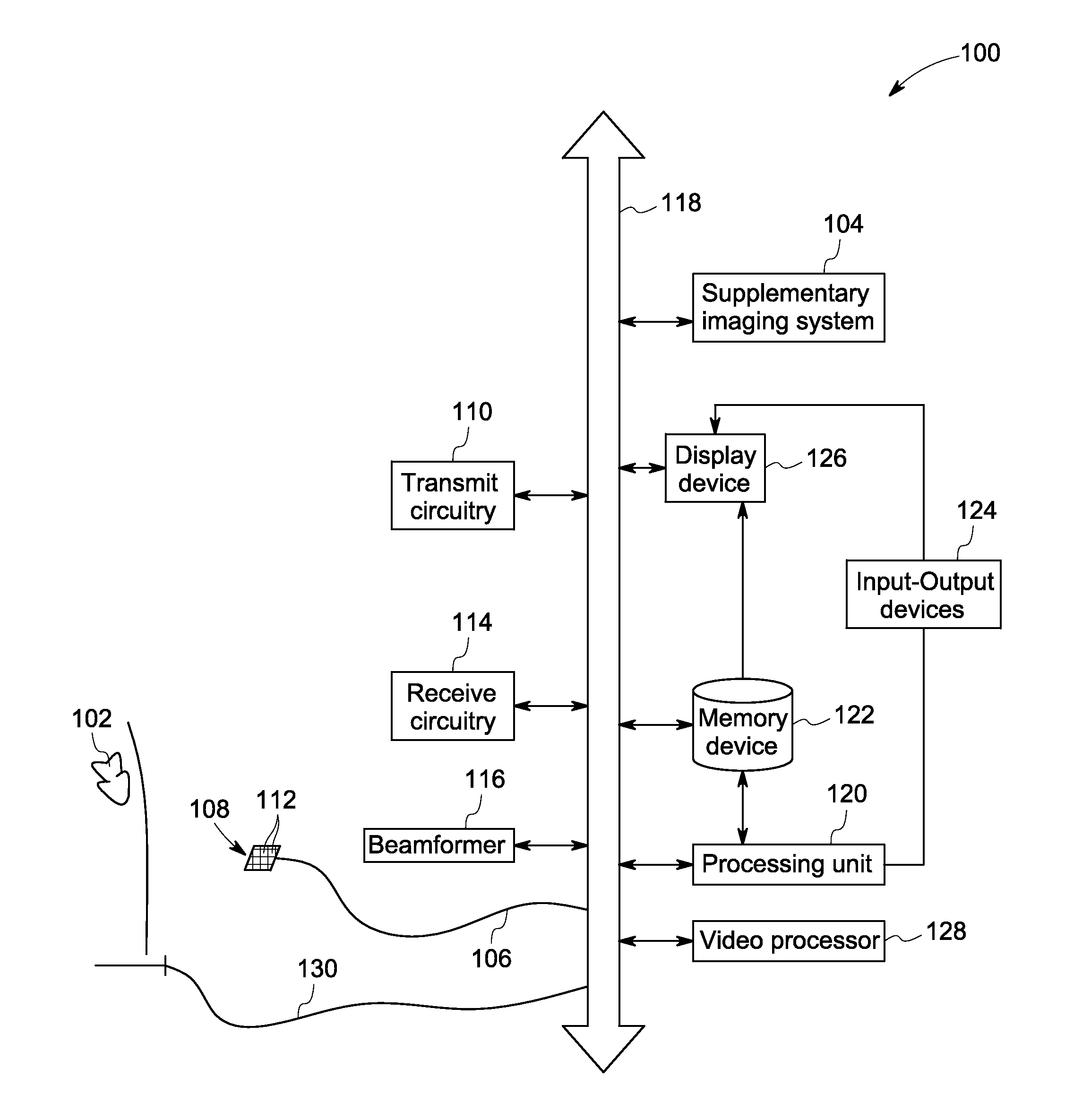
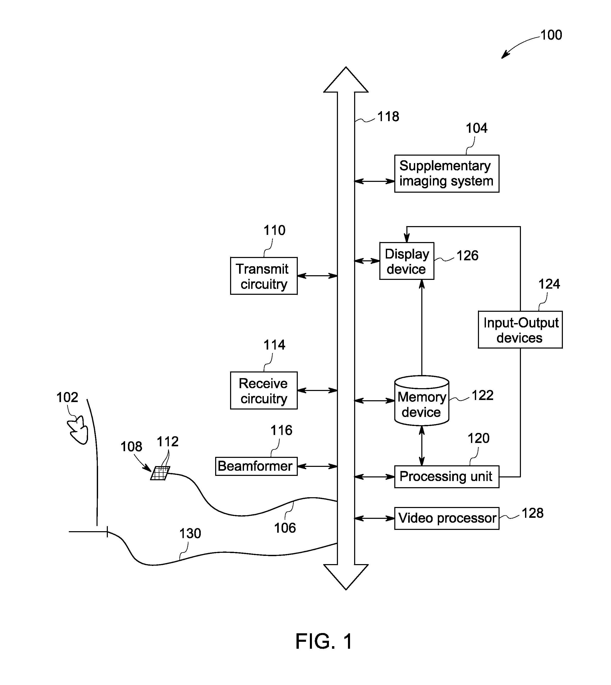
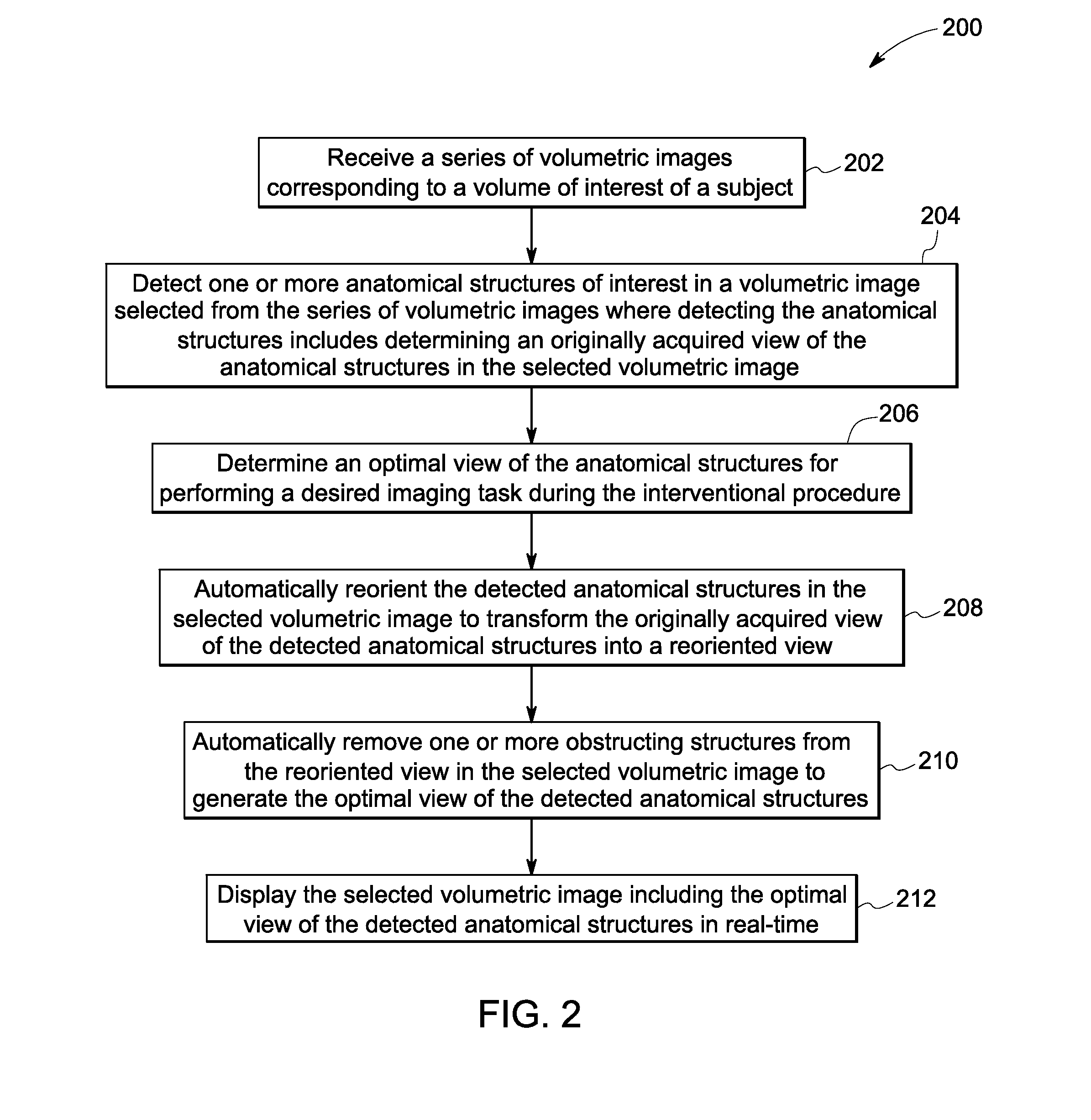
[0021]The following description presents systems and methods for optimal visualization of target anatomical structures of interest for use during interventional procedures. Particularly, certain embodiments illustrated herein describe methods and systems that are configured to automatically process a series of volumetric images to transform an originally acquired view of a target structure into a desired view that is relevant to an interventional procedure being performed. For example, a technical effect of the present disclosure is to provide automatic reorientation of the originally acquired view of the target structure such as a pulmonary vein in the cardiac region of a patient to provide a reoriented view of the target structure. Furthermore, one or more obstructing structures such as a septum may be removed from the reoriented view to provide an optimal view for ablating desired regions of the pulmonary vein. Automatic reorientation and / or removal of obstructing anatomy preclud...


PUM


Abstract

Methods and systems for imaging a subject are presented. A series of volumetric images corresponding to a volume of interest in the subject is received during an interventional procedure. One or more of anatomical structures in at least one volumetric image selected from the series of volumetric images are detected. Detecting the anatomical structures includes determining an originally acquired view of the anatomical structures in the selected volumetric image. An optimal view of the anatomical structures is determined for performing a desired imaging task during the interventional procedure. The detected anatomical structures are automatically reoriented to transform the originally acquired view of the detected anatomical structures into a reoriented view. One or more obstructing structures are automatically removed from the reoriented view to generate the optimal view of the detected anatomical structures. The selected volumetric image including the optimal view of the detected anatomical structures is displayed in real-time.

Description

BACKGROUND[0001]Embodiments of the present disclosure relate generally to interventional imaging and, more particularly, to methods and systems for optimal visualization of a target region for use in interventional procedures.[0002]Interventional techniques are widely used for managing a plurality of life-threatening medical conditions. Particularly, certain interventional techniques entail minimally invasive image-guided procedures that provide a cost-effective alternative to invasive surgery. Additionally, the minimally invasive interventional procedures minimize pain and trauma caused to a patient, thereby resulting in shorter hospital stays. Accordingly, minimally invasive transcatheter therapies have found extensive use, for example, in diagnosis and treatment of valvular and congenital heart diseases. The transcatheter therapies may be further facilitated through multi-modality imaging that aids in planning, guidance, and evaluation of procedure related outcomes and complicati...

Claims


Application Information

Patent Timeline
IPC IPC(8): A61B19/00A61B5/00A61B8/00A61B6/03A61B1/04A61B17/00G01R33/28A61B8/08
CPCA61B19/5244A61B2576/00A61B5/0035A61B8/0841A61B6/032A61B6/037A61B5/0066A61B1/04A61B17/00234A61B8/085A61B5/4887A61B8/466A61B2019/5251A61B2019/5255A61B2019/5276A61B2019/524A61B2019/5236A61B2019/5263A61B5/055G01R33/285A61B5/0044A61B5/0073A61B2576/023A61B6/466A61B6/5205A61B6/5252A61B8/12A61N7/02A61B8/5207A61B8/523G06T2207/10076G06T2207/30048A61B2017/00243G06T19/20G06T2210/41G06T2219/2016G06T2219/2021A61B18/12A61B34/20A61B2034/105A61B90/37G06T7/73G16H30/40A61B1/000094
Inventor PATWARDHAN, KEDAR ANILMILLER, JAMES VRADENBURGTIAN, TAI-PENG
Owner GENERAL ELECTRIC CO
Why Eureka
Try Eureka