Visualization of Eye Anatomy

a visualization and eye anatomy technology, applied in the field of system and method for visualizing eye anatomy, can solve the problems of poor vision or even blindness, improper formation of blood-retinal barrier, weak and permeable retinal blood vessels, etc., and achieve the effect of affordable for clinicians and patients, and convenient us

Active Publication Date: 2015-06-25
SANOVAS
View PDF8 Cites 16 Cited by
  • Summary
  • Abstract
  • Description
  • Claims
  • Application Information

AI Technical Summary

Benefits of technology

[0012]The system and method of the present invention provides a number of advantages over the known optical imaging systems. On one hand, it is inexpensive and easy to use, thereby not requiring highly specialized training of physicians and making it more affordable to clinicians and patients. On the other hand, the device of the present invention allows an examining physician to obtain a much wider field of view as compared to conventional imaging techniques, thereby making the early diagnosis of diabetic retinopathy and other eye conditions and diseases more likely. In addition, the system and method provides clinicians the ability to use one or more spectrums and one or more wavelengths to visualize the anatomy of the eye.

Problems solved by technology

Diabetic retinopathy can be a serious condition and can often lead to poor vision or even blindness if not treated timely.
These changes lead to improper formation of the blood-retinal barrier and make the retinal blood vessels become weak and more permeable.
A problem with diagnosing diabetic retinopathy is that it typically begins at the periphery of the retina, which is difficult to see, and then works its way towards the center of the retina, at which point it can be too late to treat and blindness can set in.
However, such examination sometimes does not reveal signs of retinopathy present at the periphery of the retina because of the limited field of view of these cameras.
While this technique has many advantages, the equipment is very complex and expensive, making it not easily accessible to all patients and clinics.
Further, a patient's pupil needs to be dilated during the procedure, which makes it more uncomfortable for the patient.
Again, while this technique has many advantages, the required equipment is typically extremely cumbersome and expensive.
While this device is rather simple and inexpensive, it still suffers from a number of disadvantages.
Additionally, because the camera does not utilize a lens, the quality of image of the eye anatomy obtained by the camera is fairly low.
Improper protein folding or a so-called misfolding, together with accrual of unfolded proteins, leads to the formation of disordered (amorphous) or ordered (amyloid fibril) aggregates.
However, deposition and accumulation of unfolded or misfolded proteins also occurs in other parts of the eye causing various disorders.

Method used


Image

  • Visualization of Eye Anatomy
  • Visualization of Eye Anatomy
  • Visualization of Eye Anatomy

Examples


Embodiment Construction

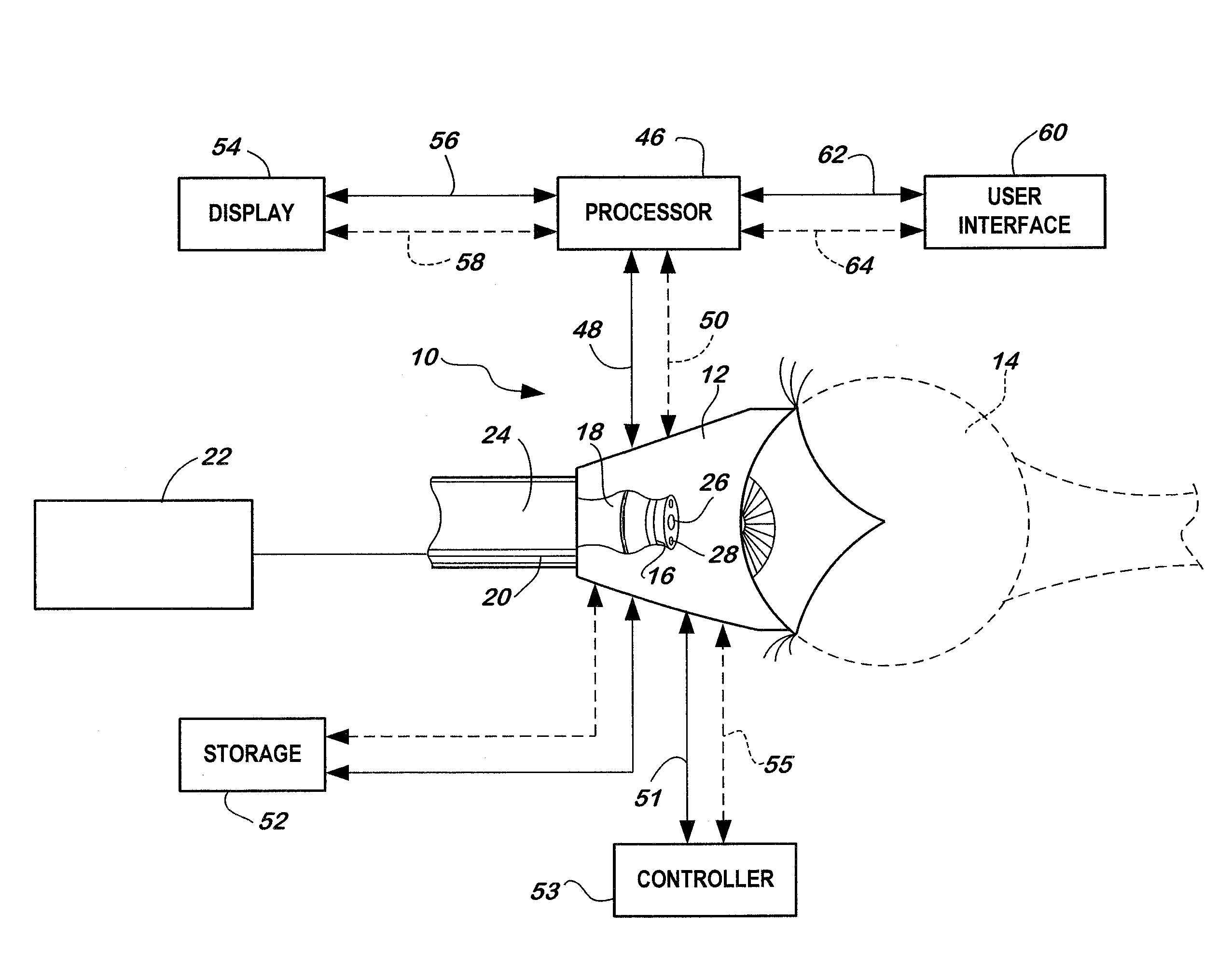
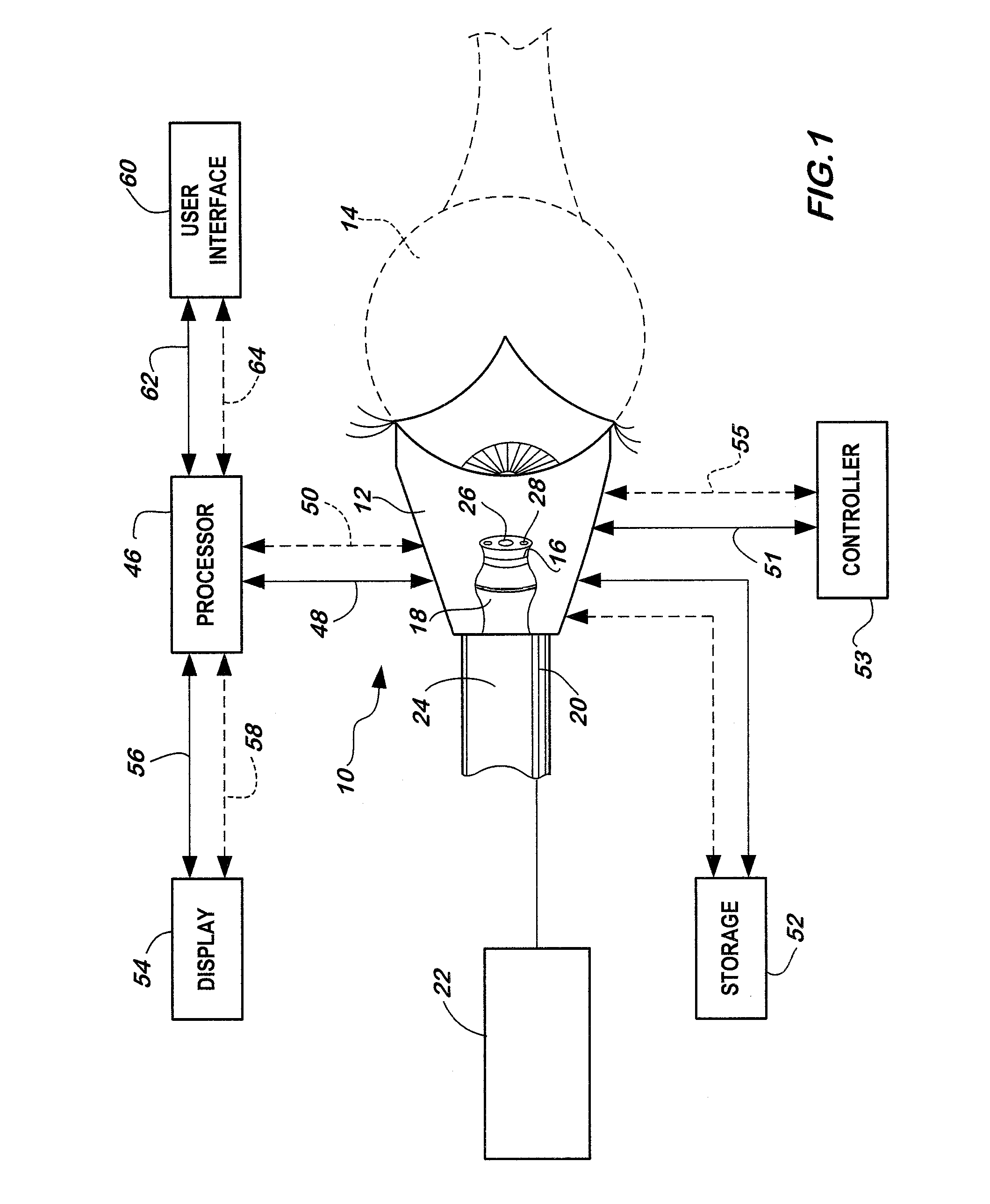
[0047]The basic components of one exemplary embodiment of a system for visualization of eye anatomy in accordance with the invention are illustrated in FIG. 1. As used in the description, the terms “top,”“bottom,”“above,”“below,”“over,”“under,”“above,”“beneath,”“on top,”“underneath,”“up,”“down,”“upper,”“lower,”“front,”“rear,”“back,”“forward” and “backward” refer to the objects referenced when in the orientation illustrated in the drawings, which orientation is not necessary for achieving the objects of the invention.

[0048]The system of the present invention utilizes a very small imaging device in combination with a vacuum-coupled contact device to image a person's eye anatomy. The imager is manually or mechanically articulatable by the physician to obtain a wide view angle of at least 180 degrees, thereby allowing examination of the periphery of the retina to detect early signs of diabetic retinopathy. It is understood that the system may also be used to image the eye anatomy for an...


PUM


Abstract

A system for visualization of eye anatomy includes at least one camera having a view vector along a first axis when in a first position, a housing to which the at least one camera is coupled, wherein the housing is configured to engage an eye of a patient such that the at least one camera is positioned adjacent the eye, and an actuator that moves the at least one camera from the first position to a second position, wherein the at least one camera, when in the second position, has a view vector along a second axis that is offset from the first axis.

Description

FIELD OF THE INVENTION[0001]The present invention relates to systems and methods for visualizing eye anatomy for diagnostic and therapeutic purposes. More specifically, the present invention relates to a system and method of visualizing eye anatomy by using a contact device together with a small articulatable imager to provide a wide angle field of view.BACKGROUND OF THE INVENTION[0002]Diabetic retinopathy is a disease characterized by damages to retina caused by complications of diabetes. The retina is a nerve layer that lines the back of the human eye. It is the part of the eye that captures the visual images and sends the images to the brain.[0003]Diabetic retinopathy can be a serious condition and can often lead to poor vision or even blindness if not treated timely. Diabetic retinopathy is typically caused by changes in retinal blood vessels, which are induced by high blood sugar levels in a diabetic patient. These changes lead to improper formation of the blood-retinal barrier...

Claims


Application Information

Patent Timeline
Patent Type & Authority Applications(United States)
IPC IPC(8): A61B3/14A61B3/113A61B3/00
CPCA61B3/14A61B3/113A61B3/0008
Inventor GERRANS, LAWRENCE J.WANG, CHARLES N.
Owner SANOVAS
Why Eureka
Try Eureka