Low-cost screening system for breast cancer detection

a screening system and low-cost technology, applied in the field of wearable equipment for breast cancer detection, can solve the problems of high cost of instruments, limited sensitivity of this technique, and unknown risk factors for subsequent cancer developmen

Inactive Publication Date: 2015-07-02
THE RES FOUND OF STATE UNIV OF NEW YORK
View PDF7 Cites 34 Cited by
  • Summary
  • Abstract
  • Description
  • Claims
  • Application Information

AI Technical Summary

Benefits of technology

[0010]According to an aspect of the present disclosure, a tumor detector is provided, which includes: a brassiere conforming to a pair of human breasts; a light emitting device configured to emit light at a plurality of emission points on the brassiere; a light sensing device configured to detect light at a plurality of detection points on the brassiere; and a controller configured to control timing of light emission at the plurality of emission points and to collect data measured by the light sensing device.
[0011]In one embodiment, the tumor detector further includes a computing means attached to the brassiere and configured to control a tomographic scan process upon the pair of human breasts. The computing means can be physically attached to the brassiere. Additionally or alternately, the computing means can be configured to generate at least one tomographic image, wherein the at least one tomographic image includes at least one of a two-dimensional map and a three-dimensional map of human breasts.

Problems solved by technology

While effective for the detection of most cancers of the breast, the sensitivity of this technique is limited by small sized tumors, especially in women with dense breasts.
Also, its use of ionizing sources is a known risk factor for subsequent cancer development, especially in women under age 50.
Still another consideration impacting its use is the cost of the instrumentation.
Today digital x-ray mammography systems typically cost several hundred thousand dollars and require considerable skill in the interpretation of the resultant images.
Other imaging methods are available, but these either lack the sensitivity required of screening methods, are operator dependent (e.g., ultrasound imaging) or employ costly instrumentation (e.g., magnetic resonance imaging (MRI)).
), an elementary challenge of the diffuse optical method in general, is its low spatial resolution.
As a consequence, the performance of the method for small tumor detection is notably reduced compared to the structural imaging methods.
While potentially promising, the need for high density sensing renders the required instrumentation bulky and costly.
Whereas imaging methods in their various forms are often successful in tumor detection, in all cases their reliance on detection of a focal contrast feature strongly limits their sensitivity to small tumors.

Method used


Image

  • Low-cost screening system for breast cancer detection
  • Low-cost screening system for breast cancer detection
  • Low-cost screening system for breast cancer detection

Examples


embodiment

Preferred Embodiment

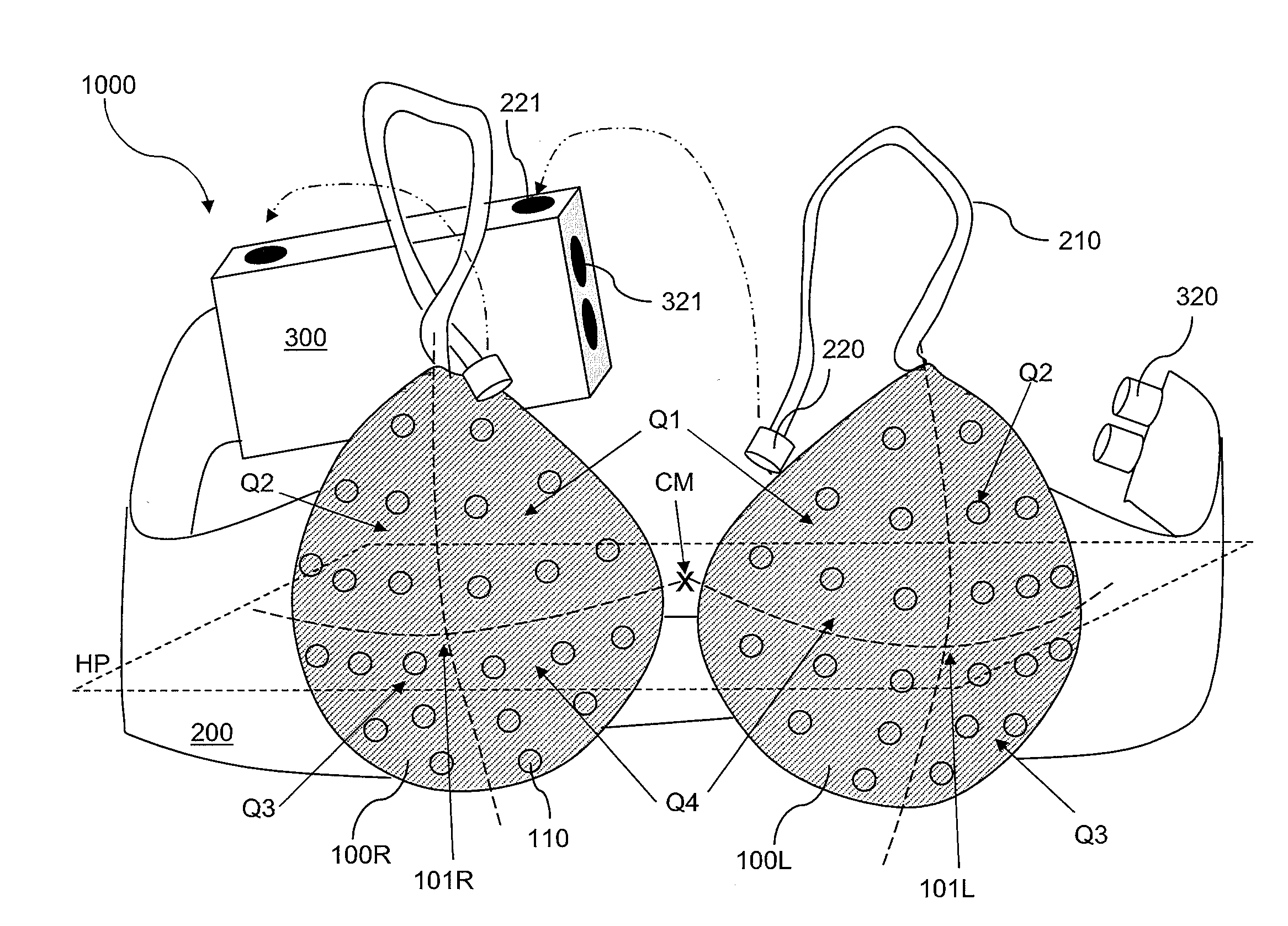
[0041]Apart from the clinical diagnostic sensitivity required to support a screening method, its utility in a home or similarly less supervised environment requires access to simplified, low-cost equipment. Illustrated in FIG. 4 is an exemplary tumor detector according to an embodiment of the present disclosure. A limited sensing array is bilaterally incorporated into a dark colored brassiere that conforms to the appropriate chest and breast size for the individual undergoing the optical examination. As used herein, a “brassiere” refers to any structure that a normal female human being can wear around her breast in a manner that covers both of her breasts without a substantial risk of disengagement of the structure from her breasts unless consciously disengaged. The brassier may be portable, which means that the brassier may be taken to any location without regard to mechanical connection or support that is available only at specific locations. The brassier can b...


PUM


Abstract

A portable and wearable tumor detector includes a brassier and devices for enabling optical tomography in a non-clinical setting. Light emitting devices and light sensing devices are provided on the brassier, and a controller for performing a tomographic scanning is attached to the brassier. A computing means and a communication means may be provided to generate at least one tomographic image. Each of the two breasts under examination can be employed as a reference structure for generating a tomographic image for the other of the two breasts, thereby providing a self-referencing image generation mechanism. The images and/or data can be reviewed by the subject of the tomographic scanning or by a medical professional. The tomographic scanning can be performed at any location if provided with a portable power supply system.

Description

CROSS-REFERENCE TO RELATED APPLICATIONS[0001]This application claims the benefit of U.S. Provisional Application No. 61 / 674,029, filed on Jul. 20, 2012.STATEMENT REGARDING FEDERALLY SPONSORED RESEARCH OR DEVELOPMENT[0002]This invention was made with government support under Contract No. R41CA096102 awarded by the National Institute of Health, Contact No. DAMD017-03-C-0018 awarded by U.S. Army, Contract No. NYSTAR-TIPP C020041 awarded by New York State Foundation for Science, Technology, and Innovation—Technology Transfer Incentive Program, Contract No. IMG0403022 to Susan G. Komen Foundation, and a grant to Brooklyn Hospital Center from New York State Department of Health through Empirical Clinical Research Investigator Program. The federal government of the United States has certain rights in the invention.FIELD OF THE INVENTION[0003]The present invention relates to a wearable apparatus for breast cancer detection and a method for operating the same.BACKGROUND OF THE INVENTION[0004...

Claims


Application Information

Patent Timeline
Patent Type & Authority Applications(United States)
IPC IPC(8): A61B5/00
CPCA61B5/0091A61B5/0073A61B5/7282A61B5/742A61B5/0013A61B5/6804
Inventor BARBOUR, RANDALL L.AL ABDI, RABAH M.GRABER, HARRY
Owner THE RES FOUND OF STATE UNIV OF NEW YORK
Why Eureka
Try Eureka