
Fully integrated, disposable tissue visualization device

Active Publication Date: 2015-07-16
TRICE MEDICAL
View PDF22 Cites 27 Cited by
  • Summary
  • Abstract
  • Description
  • Claims
  • Application Information

AI Technical Summary

Benefits of technology

The patent describes an imaging device that can be used to accurately position a distal end of an elongated body, such as within a joint capsule, for the diagnosis and treatment of injuries or conditions. The device provides visual and tactile feedback to the operator and allows for reliable delivery of therapeutic media into a joint without the need for other diagnostic procedures that may be less accurate or require a longer wait period.

Problems solved by technology

Traditional surgical procedures, both therapeutic and diagnostic, for pathologies located within the body can cause significant trauma to the intervening tissues.
These procedures often require a long incision, extensive muscle stripping, prolonged retraction of tissues, denervation and devascularization of tissue.
Such procedures can require operating room time of several hours followed by several weeks of post-operative recovery time due to the destruction of tissue during the surgical procedure.
In some cases, these invasive procedures lead to permanent scarring and pain that can be more severe than the pain leading to the surgical intervention.
However, proper visualization of an internal tissue site can be expensive and time-consuming to schedule, such as when magnetic resonance imaging (MRI) is required.
Further, other modes of imaging can potentially damage the tissue site, leading to poor diagnosis and extended recovery time.

Method used


Image

  • Fully integrated, disposable tissue visualization device
  • Fully integrated, disposable tissue visualization device
  • Fully integrated, disposable tissue visualization device

Examples


Embodiment Construction

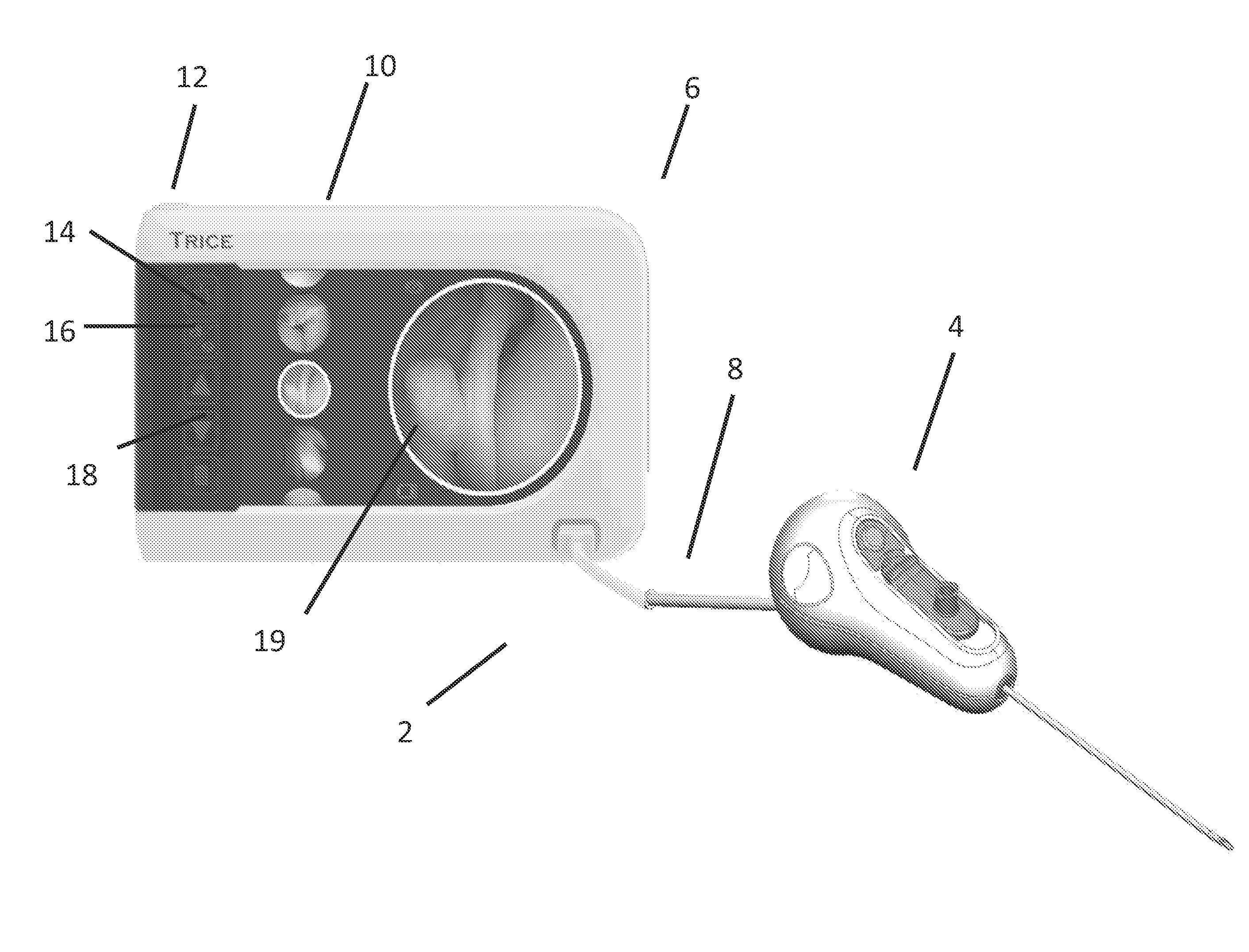
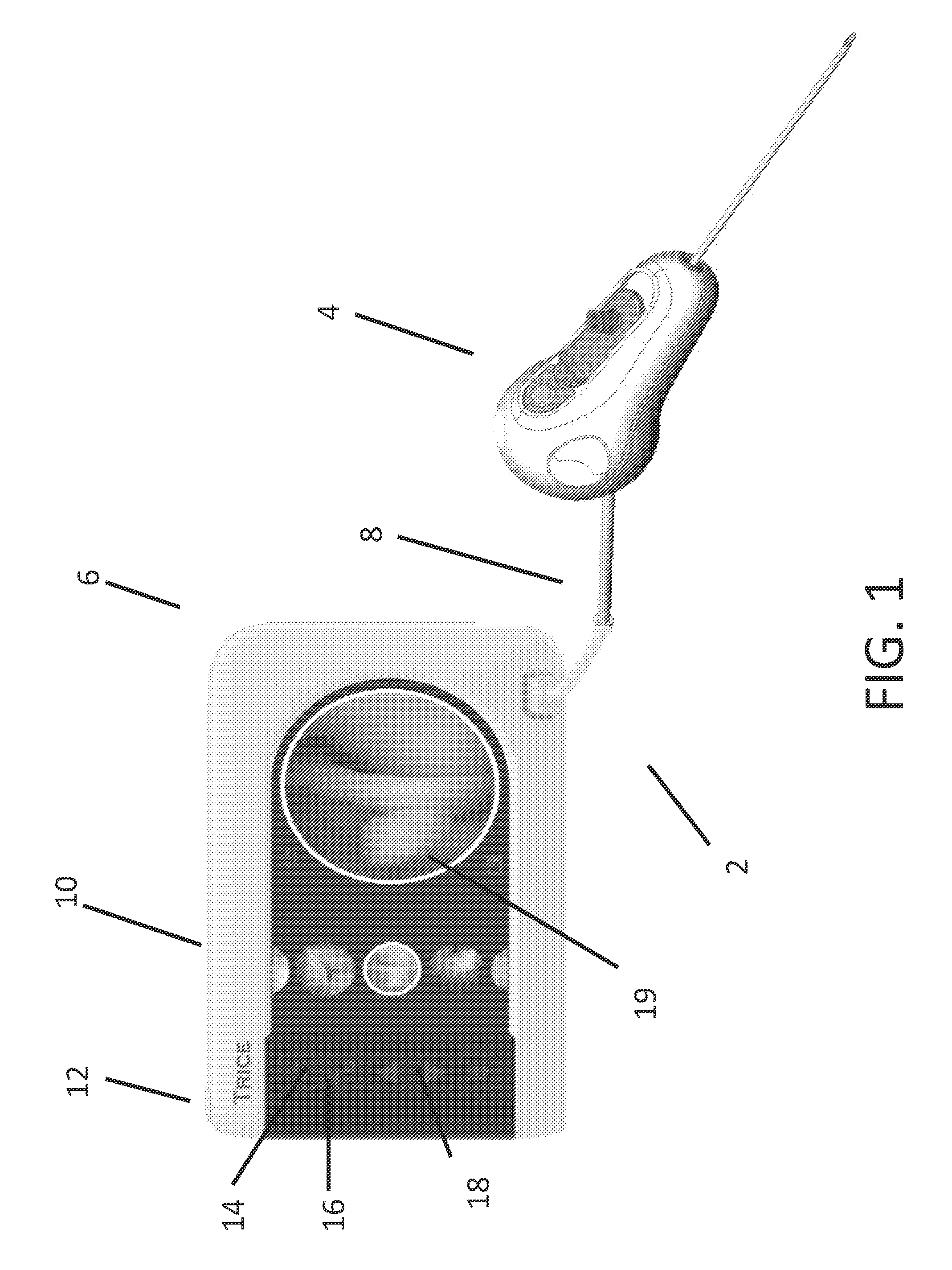
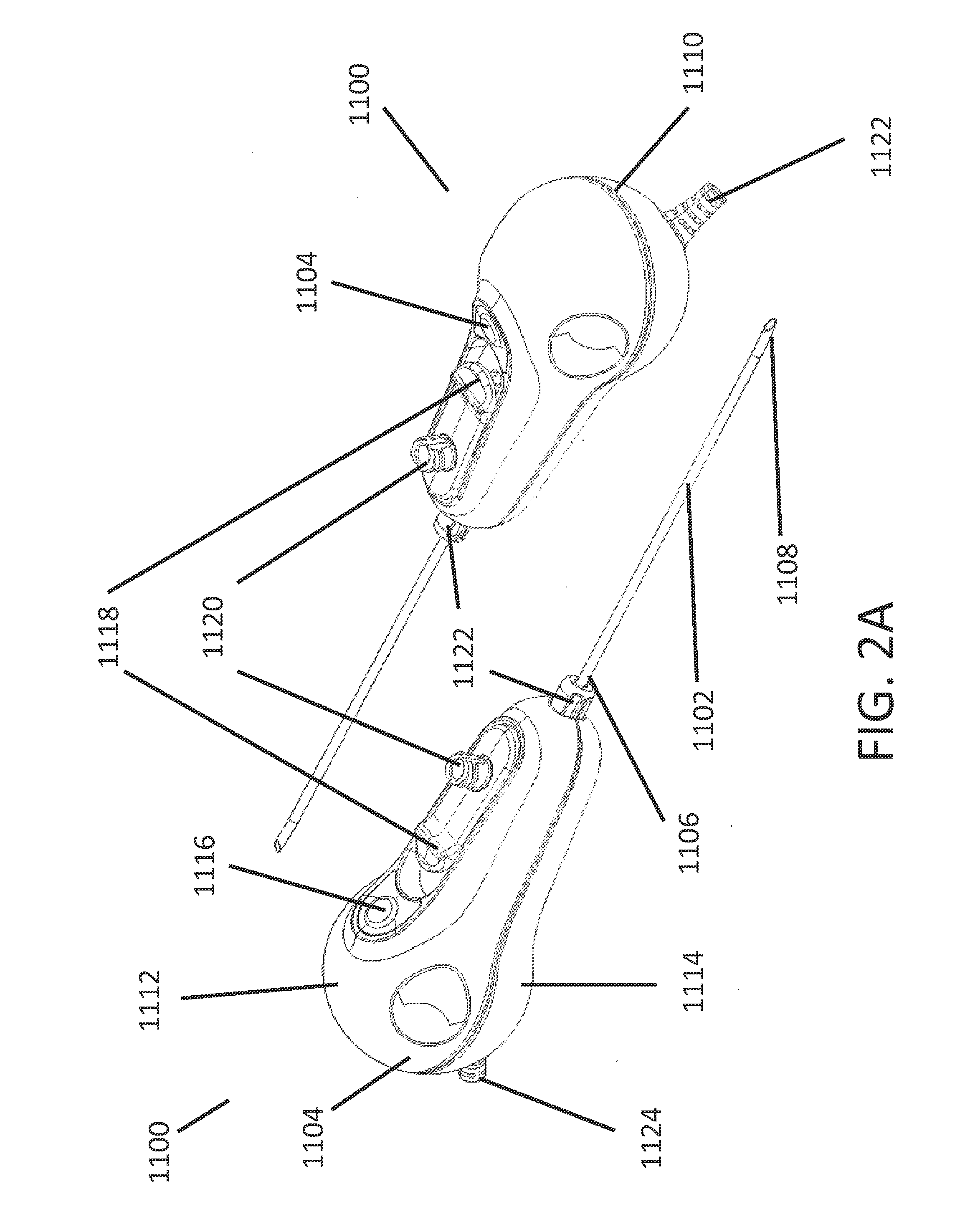
[0046]Embodiments disclosed in this section or elsewhere in this application relate to minimally invasive tissue visualization and access systems and devices. Also provided are methods of using the systems in imaging applications, as well as kits for performing the methods.

[0047]Before the present invention is described in greater detail, it is to be understood that this invention is not limited to particular embodiments described, as such may, of course, vary. It is also to be understood that the terminology used herein is for the purpose of describing particular embodiments only, and is not intended to be limiting, since the scope of the present invention will be limited only by the appended claims.

[0048]Where a range of values is provided, it is understood that each intervening value between the upper and lower limit of that range and any other stated or intervening value in that stated range, is encompassed within the invention. The upper and lower limits of these smaller ranges...


PUM


Abstract

The present invention relates to a fully integrated sterilizable one time use disposable tissue visualization device and methods for using such devices. Preferred embodiments of the invention facilitate the visualization of an internal tissue site while causing a minimum of damage to the surrounding tissue. Further preferred embodiments may allow for the delivery of fluids and other treatment to an internal tissue site.

Description

CROSS-REFERENCE TO RELATED APPLICATIONS[0001]This application claims the priority benefit under 35 U.S.C. §119(e) of U.S. Provisional Application No. 61 / 926,578, filed Jan. 13, 2014, the entirety of which is hereby incorporated by reference herein.BACKGROUND OF THE INVENTION[0002]1. Field of the Invention[0003]This application describes embodiments of apparatuses, methods, and systems for the visualization of tissues.[0004]2. Description of the Related Art[0005]Traditional surgical procedures, both therapeutic and diagnostic, for pathologies located within the body can cause significant trauma to the intervening tissues. These procedures often require a long incision, extensive muscle stripping, prolonged retraction of tissues, denervation and devascularization of tissue. Such procedures can require operating room time of several hours followed by several weeks of post-operative recovery time due to the destruction of tissue during the surgical procedure. In some cases, these invasi...

Claims


Application Information

Patent Timeline
IPC IPC(8): A61B1/00A61B17/34A61B1/07
CPCA61B1/00103A61B17/3468A61B1/00066A61B1/07A61B1/00087A61B1/00098A61B1/0011A61B1/00163A61B1/012A61B1/00045A61B1/00114A61B1/002A61B1/045A61B1/0684A61B17/34A61B2017/0023A61B1/00016A61B1/0002A61B1/00108A61B1/00154A61B1/015A61B1/0676A61B1/3135A61B1/317A61B17/3415A61B2017/3456
Inventor KIENZLE, RICHARD A.WASHBURN, II, RICHARD H.BRIGANTI, RICHARD T.
Owner TRICE MEDICAL
Learn More