System, method and computer-accessible medium for rapid real-time cardiac magnetic resonance imaging utilizing synchronized cardio-respiratory sparsity

a real-time cardiac and magnetic resonance imaging technology, applied in the field of magnetic resonance imaging, can solve the problems of limiting temporal sparsity, introducing spatial blurring due to interpolation errors, and affecting the performance of breath-hold cine mri in patients with impaired breath-hold capability

Active Publication Date: 2015-10-29
NEW YORK UNIV
View PDF3 Cites 11 Cited by
  • Summary
  • Abstract
  • Description
  • Claims
  • Application Information

AI Technical Summary

Benefits of technology

The patent describes a system, method, and computer-accessible medium for generating images of tissues, such as cardiac and respiratory tissues, using magnetic resonance imaging (MRI) information. The MRI information is received and parsed into two dimensions, which can be motion-related dimensions. The system uses a golden-angle radial sampling procedure to sort and synchronize the MRI information. The resulting images can be generated based on the parsed information using a joint multi-coil compressed sensing procedure. The system can also use a residual streaking artifact removal technique to improve image quality. The patent also mentions that the system can use other types of imaging information, such as positron emission tomography, computed tomography, or single-photon emission computed tomography. Overall, the patent provides a technical solution for generating high-quality images of tissues using MRI information.

Problems solved by technology

However, the performance of breath-hold cine MRI can be degraded in patients with impaired breath-hold capability, due to failure to synchronize cardiac cycles at different respiratory states.
In order to minimize the influence from respiration, a deformable registration framework has been incorporated into reconstruction for respiratory motion correction (see, e.g., References 2 and 3); however it ran the risk of introducing spatial blurring due to interpolation errors.
However, the superposition of respiratory and cardiac motion limits temporal sparsity.
Evaluation of myocardial function with MRI can be challenging in patients with arrhythmias, such as premature ventricular contractions (“PVCs”) or atrial fibrillation, due to the difficulty of synchronizing disparate cardiac cycles.
However, conventional temporal compressed sensing does not account for respiratory motion or arrhythmias, and thus only moderate performance can be achieved in these cases.

Method used


Image

  • System, method and computer-accessible medium for rapid real-time cardiac magnetic resonance imaging utilizing synchronized cardio-respiratory sparsity
  • System, method and computer-accessible medium for rapid real-time cardiac magnetic resonance imaging utilizing synchronized cardio-respiratory sparsity
  • System, method and computer-accessible medium for rapid real-time cardiac magnetic resonance imaging utilizing synchronized cardio-respiratory sparsity

Examples


Embodiment Construction

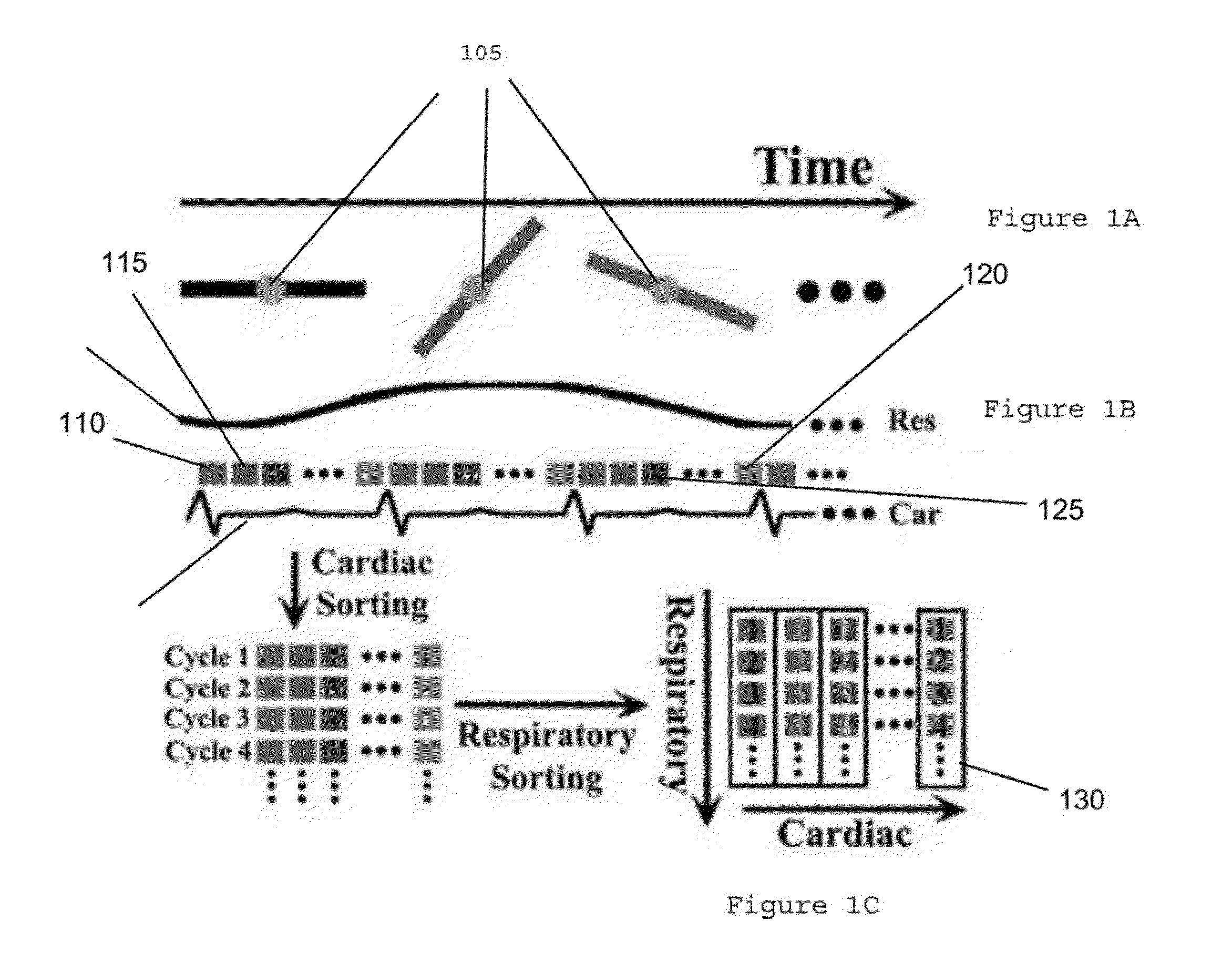
[0006]An exemplary system, method and computer-accessible medium can be provided for generating an image(s) of a tissue(s) that can include, for example, receiving magnetic resonance imaging information regarding the tissue(s) can based on a golden-angle radial sampling procedure, sorting and synchronizing the MRI information into at least two dimensions, which can be motion-related dimensions, and generating the image(s) of the tissue(s) based on the sorted and synchronized information. The tissue(s) can include cardiac tissue and respiratory tissues (e.g., respiratory-affected tissue). The MRI information can include both motion of the cardiac tissue and motion of the respiratory tissue.

[0007]In some exemplary embodiments of the present disclosure, the dimensions can be two separated dimensions. The image(s) can be generated based on a compressed sensing procedure, which can be a joint multi-coil compressed sensing procedure. The compressed sensing procedure can optionally be perf...


PUM


Abstract

An exemplary system, method and computer-accessible medium can be provided for generating an image(s) of a tissue(s) that can include, for example, receiving magnetic resonance imaging information regarding the tissue(s) based on a golden-angle radial sampling procedure, sorting and synchronizing the MRI information into at least two dimensions, and generating the image(s) of the tissue(s) based on the sorted and synchronized information. The tissue(s) include cardiac tissue and respiratory-affected tissue. The MRI information can include a motion of the cardiac tissue and a motion of the respiratory tissue.

Description

CROSS-REFERENCE TO RELATED APPLICATION(S)[0001]This application relates to and claims priority from U.S. Patent Application No. 61 / 984,364, filed on Apr. 25, 2014, the entire disclosure of which is incorporated herein by reference.FIELD OF THE DISCLOSURE[0002]The present disclosure relates generally to magnetic resonance imaging (“MRI”), and more specifically, to exemplary embodiments of an exemplary system, method and computer-accessible medium for rapid real-time cardiac MRI utilizing synchronized cardio-respiratory sparsity.BACKGROUND INFORMATION[0003]Breath-hold balanced-Steady-State Free Precession (b-“SSFP”) cine imaging can be considered the gold standard for evaluating myocardial function in MRI. (See, e.g., Reference 1). However, the performance of breath-hold cine MRI can be degraded in patients with impaired breath-hold capability, due to failure to synchronize cardiac cycles at different respiratory states. In order to minimize the influence from respiration, a deformabl...

Claims


Application Information

Patent Timeline
Patent Type & Authority Applications(United States)
IPC IPC(8): G01R33/48G01R33/567A61B5/05G01R33/565
CPCG01R33/4824G01R33/56509A61B5/05G01R33/4828G01R33/5673A61B5/7207A61B5/0044A61B5/055A61B5/7289G01R33/56325
Inventor AXEL, LEONFENG, LLOTAZO, RICARDOSODICKSON, DANIEL
Owner NEW YORK UNIV
Why Eureka
Try Eureka