Noninvasive 4-D time-resolved dynamic magnetic resonance angiography

a dynamic magnetic resonance and time-resolved technology, applied in the field of magnetic resonance imaging, can solve the problems of invasive dsa procedure, difficult to derive quantitative hemodynamic information using either dsa and/or ce-dmra procedures, and associated risks of allergic and other adverse reactions, and achieve high spatial and temporal resolution, adequate snr, and high degree of efficiency and flexibility

Active Publication Date: 2019-11-12
THE TRUSTEES OF THE UNIV OF PENNSYLVANIA +1
View PDF12 Cites 0 Cited by
  • Summary
  • Abstract
  • Description
  • Claims
  • Application Information

AI Technical Summary

Benefits of technology

[0016]However, compared to the standard arterial spin labeling (ASL) methods that offer a single physiological parameter (blood flow) at a single post-labeling delay time, the invention provides 4D dynamic information of blood flow with high temporal resolution and spatial resolution, as well as quantification of multiple hemodynamic parameters such as blood flow, blood volume and mean transit time. The use of accelerated imaging methods and enhanced / prolonged labeling bolus produces superior results compared to existing imaging methods due to higher imaging speed, spatio-temporal resolution and signal-to-noise ratio. The high temporal resolution (50-100 ms) of the technique allows the scan to be repeated as often as required during the same session.
[0020]In one embodiment, a dynamic radial acquisition with a golden angle view increment is used with the 4D dMRA method because of its high degree of efficiency and flexibility for retrospective dynamic image reconstruction. In another embodiment, a 3D stack-of-stars golden-angle radial acquisition in conjunction with temporal filtering strategies (k-space weighted image contrast or KWIC) is used to achieve ultrafast 4D dMRA with high spatial and temporal resolution, adequate SNR, and high temporal fidelity.
[0021]According to one aspect of the invention, a method for magnetic resonance angiography is provided that has high spatial resolution for depicting vascular architecture, as well as high temporal resolution for visualizing dynamic blood flow patterns.

Problems solved by technology

The DSA procedure, however, is invasive and requires the use of ionizing radiation (X-rays) as well as the injection of iodinated contrast media for imaging that has the associated risks of allergic and other adverse reactions.
In addition, it remains challenging to derive quantitative hemodynamic information using either the DSA and / or the CE-dMRA procedures.
However, past attempts to formulate schemes that reduce the time of acquisition and the amount of contrast exposure while increasing spatial resolution are usually at the expense of SNR and image quality.

Method used


Image

  • Noninvasive 4-D time-resolved dynamic magnetic resonance angiography
  • Noninvasive 4-D time-resolved dynamic magnetic resonance angiography
  • Noninvasive 4-D time-resolved dynamic magnetic resonance angiography

Examples


example 1

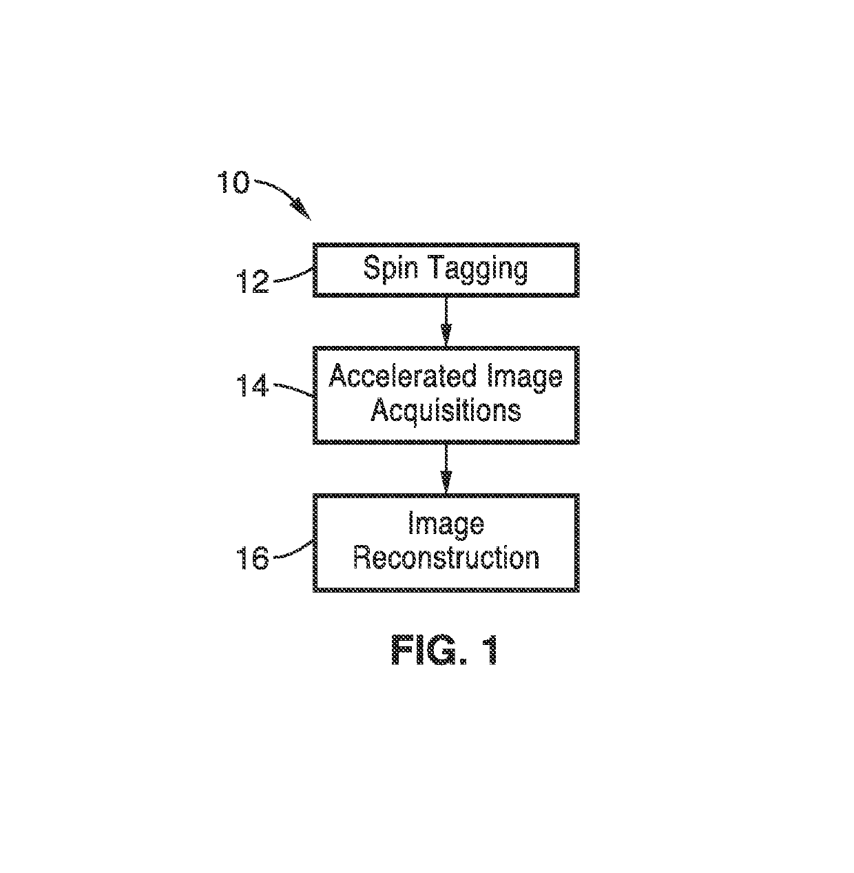
[0049]In order to demonstrate the functionality of the spin labeling aspects of the invention, a FISP-based spin tagging with alternating radiofrequencies (STAR) labeling sequence was evaluated and the spatial and temporal resolution of the method was optimized in eight healthy volunteers. In another six healthy volunteers, the contrast-to-noise ratio (CNR) and signal-to-noise ratio (SNR) of the STAR based dynamic MR angiography images were compared with those acquired by using a standard Look-Locker echo-planar technique by using the Wilcoxon signed rank test. Finally, one patient with an arteriovenous malformation (AVM) was studied using this technique.

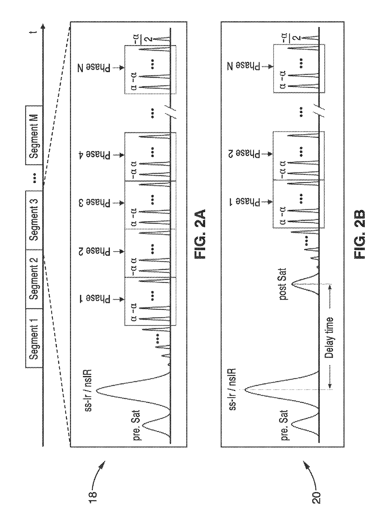
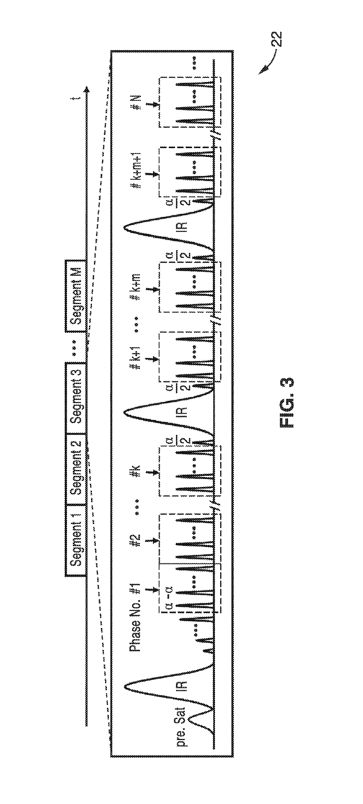
[0050]As shown in the pulse sequence diagrams of FIG. 2A and FIG. 2B, immediately following a section-selective or nonselective hyperbolic secant inversion pulse, a train of 20 dummy radiofrequency pulses with Kaiser Bessel window ramp flip angles (i.e., α / 21, −2α / 21, 3α / 21, . . . , 20α / 21) was applied to minimize transient signal o...

example 2

[0057]To further demonstrate the invention, two experiments were conducted with eight healthy volunteers (24.6±3.6 yrs, 3 males) on a Siemens TIM Trio 3T scanner. In the first experiment, a three-bolus STAR sequence was implemented with the time interval of 210, 315, 420 and 525 ms between inversion pulses respectively. The tagging was applied to an 80 mm slab inferior to the image slab with a 20 mm gap. The rest parameters were: FOV=220×165 mm2, resolution=1×1×1.5 mm3, rate-2 GRAPPA; a 3D slab of 40 slices with 1.5 mm thickness, 22 phases from 150 to 2370 ms with a step of 105 ms was acquired within a total scan time of 7 minutes.

[0058]The second experiment made a comparison of the optimized multi-bolus dMRA with standard single-bolus PASL and pCASL based dMRA. The phase interval of 4 (420 ms) between inversion pulses was chosen for multi-bolus dMRA, with the rest parameters identical to the protocol used in the first experiment. For comparison, a standard single-bolus STAR sequenc...

example 3

[0061]The ultrafast 4D dMRA methods were further demonstrated with an embodiment employing a 3D stack-of-stars golden-angle radial acquisition in conjunction with temporal filtering strategies of k-space weighted image contrast (KWIC) to achieve 4D dMRA with high spatial and temporal resolution, adequate SNR, and high temporal fidelity. Dynamic radial acquisition with a golden angle view increment is ideally suited for 4D dMRA, given its high degree of efficiency and flexibility for retrospective dynamic image reconstruction.

[0062]The pulse sequence that was used consisted of a continuous 3D TrueFISP readout following either slice-selective or non-selective inversion pulses. A dynamic 3D radial stack-of-stars sampling with an in-plane view angle increment of θg=111.25° (golden angle) was utilized as illustrated in FIG. 4. A 3D radial stack-of-stars sampling was achieved by taking multiple shots, where each shot represented a slice-encoding step. The imaging parameters were as follow...


PUM


Abstract

A method for non-contrast enhanced 4D time resolved dynamic magnetic resonance angiography using arterial spin labeling of blood water as an endogenous tracer and a multiphase balanced steady state free precession readout is presented. Imaging can be accelerated with dynamic golden angle radial acquisitions and k-space weighted imaging contrast (KWIC) image reconstruction and it can be used with parallel imaging techniques. Quantitative tracer kinetic models can be formed allowing cerebral blood volume, cerebral blood flow and mean transit time to be estimated. Vascular compliance can also be assessed using 4D dMRA by synchronizing dMRA acquisitions with the systolic and diastolic phases of the cardiac cycle.

Description

CROSS-REFERENCE TO RELATED APPLICATIONS[0001]This application is a 35 U.S.C. § 111(a) continuation of PCT international application number PCT / US2013 / 071590 filed on Nov. 25, 2013, incorporated herein by reference in its entirety, which claims priority to, and the benefit of, U.S. provisional patent application Ser. No. 61 / 731,278 filed on Nov. 29, 2012, incorporated herein by reference in its entirety. Priority is claimed to each of the foregoing applications.[0002]The above-referenced PCT international application was published as PCT International Publication No. WO 2014 / 085288 on Jun. 5, 2014, which publication is incorporated herein by reference in its entirety.STATEMENT REGARDING FEDERALLY SPONSORED RESEARCH OR DEVELOPMENT[0003]This invention was made with Government support under MH080892, awarded by the National Institutes of Health. The Government has certain rights in this invention.INCORPORATION-BY-REFERENCE OF MATERIAL SUBMITTED IN A COMPUTER PROGRAM APPENDIX[0004]Not Ap...

Claims


Application Information

Patent Timeline
Patent Type & Authority Patents(United States)
IPC IPC(8): G01R33/48A61B5/026G01R33/563A61B5/055G01R33/56G01R33/561
CPCG01R33/4806G01R33/4828A61B5/0263A61B5/055G01R33/4818G01R33/5635G01R33/56333G01R33/5611G01R33/5614G01R33/4824G01R33/5601
Inventor WANG, DANNY J JYAN, LIRONGSONG, HEE KWON
Owner THE TRUSTEES OF THE UNIV OF PENNSYLVANIA
Why Eureka
Try Eureka