
Standalone annotations of axial-view spine images

a spine and axial view technology, applied in the field of labeling radiology images, can solve the problems of difficult difficult for radiologists to determine the identity of a particular vertebra, and not always available sagittal images for every patien

Active Publication Date: 2015-11-12
LONDON HEALTH SCI CENT RES +1
View PDF3 Cites 9 Cited by
  • Summary
  • Abstract
  • Description
  • Claims
  • Application Information

AI Technical Summary

Benefits of technology

The patent text describes a system and method for annotating axial spine images without the need for additional views or external training. The system can be used with different imaging modalities and acquisition protocols. The technical effect of the system and method is that it provides a more efficient and automated way to annotate spine images, which can save time and improve the accuracy of the annotations.

Problems solved by technology

Generating labels in a manual fashion is tedious, subjective, and time-consuming especially because the number of 2D slices in a series can be very high, with up to 100 images per axial series.
Furthermore, it can be difficult for radiologists to determine the identity of a particular vertebra in an axial slice of the spine, because adjacent vertebrae have very similar appearances.
First, sagittal images are not always available for every patient (i.e., only the axial view may be available) while in other cases the two scans might be acquired at different time points.
In such cases, when deformations between sagittal and axial images occur because of patient repositioning, cross-referencing may not be reliable.
Therefore, localizing the spinal structures in different views becomes challenging (even for an experienced radiologist).
Such algorithms work very well on data sets that closely match the training data, but require adjustment / retraining for different data sets or if the imaging modality and / or acquisitions protocol are altered (e.g., an algorithm that is trained and built for Computerized Tomography (CT) images may not perform well on Magnetic Resonance (MR) data).
This impedes the use of these algorithms in routine clinical practices, where a particular disorder might be analyzed radiologically using several different imaging modalities / protocols with widely variable imaging parameters.

Method used


Image

  • Standalone annotations of axial-view spine images
  • Standalone annotations of axial-view spine images
  • Standalone annotations of axial-view spine images

Examples


Embodiment Construction

[0016]In the following detailed description, reference is made to the accompanying drawings that form a part hereof, and in which is shown by way of illustration specific examples that may be practiced. These examples are described in sufficient detail to enable one skilled in the art to practice the subject matter, and it is to be understood that other examples may be utilized and that logical, mechanical, electrical and other changes may be made without departing from the scope of the subject matter of this disclosure. The following detailed description is, therefore, provided to describe an exemplary implementation and not to be taken as limiting on the scope of the subject matter described in this disclosure. Certain features from different aspects of the following description may be combined to form yet new aspects of the subject matter discussed below.

[0017]When introducing elements of various embodiments of the present disclosure, the articles “a,”“an,”“the,” and “said” are i...


PUM


Abstract

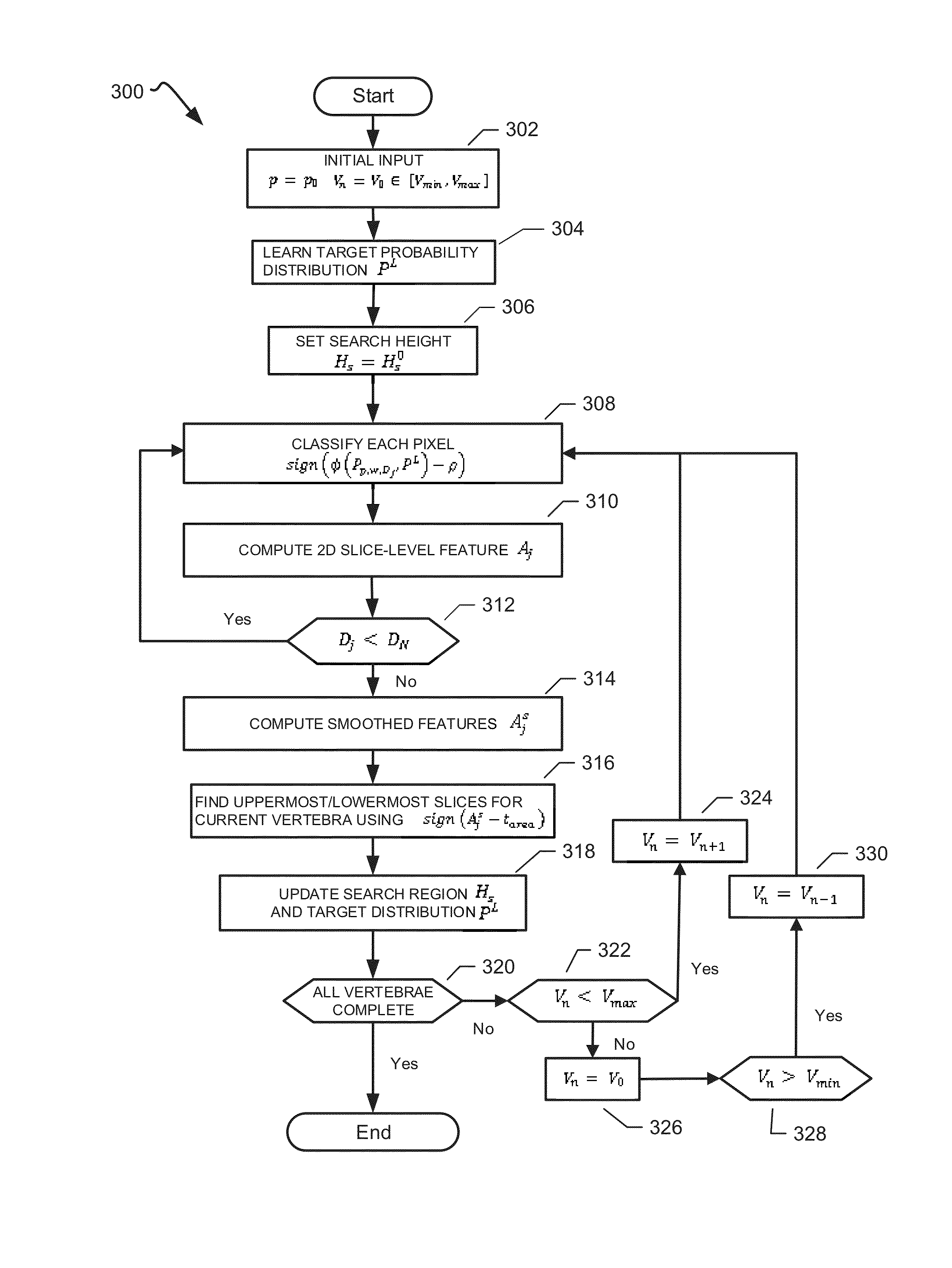
Systems, methods and computer program products to annotate axial-view spine images with the desired characteristics of not requiring additional views of the spine or cross-referencing features are provided. In one aspect, the disclosed method does not require external training from a manually-labeled data set while being applicable to different imaging modalities and acquisition protocols. In one aspect, the disclosed method achieves near real-time results using integral kernels when implemented on a Graphics Processing Unit.

Description

FIELD OF DISCLOSURE[0001]The present disclosure relates to labeling radiology images, and more particularly to systems, methods and computer program products to label radiology images using the axial view.BACKGROUND[0002]The statements in this section merely provide background information related to the disclosure and may not constitute prior art.[0003]The annotations (i.e., labels) of the vertebrae and inter-vertebral discs in axial spine Magnetic Resonance Image (MRI) images (i.e., slices) are essential for further diagnosis of various spine disorders. For instance, in MRI, annotating the axial-view slices facilitates the quantification and level-based reporting of common inter-vertebral disc displacements such as protrusion, extrusion, and bulging.[0004]Generating labels in a manual fashion is tedious, subjective, and time-consuming especially because the number of 2D slices in a series can be very high, with up to 100 images per axial series. Furthermore, it can be difficult for...

Claims


Application Information

Patent Timeline
IPC IPC(8): G06T11/60G06K9/62G06T7/00
CPCG06T11/60G06T7/0012G06K9/6277G06T2207/30012G06T2207/10116G06T2207/20076G06T2207/20081G06K9/6215A61B6/032A61B6/468A61B6/505A61B6/5211G06T2207/10088G06T7/11A61B5/0033G06F18/22G06F18/2415
Inventor BEN AYED, ISMAILMILES, BRANDONGARVIN, GREGORY
Owner LONDON HEALTH SCI CENT RES
Learn More