Systems and Methods for Percutaneous Access, Stabilization and Closure of Organs

a technology of percutaneous access and organs, applied in the direction of staples, nails, diagnostics, etc., can solve the problems of inconvenient operation, inconvenient operation, and inconvenient operation of percutaneous access and other problems, to achieve the effect of improving the ease and safety of conduit insertion, facilitating the operation and facilitating the removal and retrieval

Inactive Publication Date: 2015-12-03
APICA CARDIOVASCULAR
View PDF6 Cites 6 Cited by
  • Summary
  • Abstract
  • Description
  • Claims
  • Application Information

AI Technical Summary

Benefits of technology

[0016]In certain embodiments, the invention provides a tissue sealing device comprising a tissue attaching device having an outer base ring defining an opening therethrough and a distally projecting tissue attachment coil defining a first pitch region and distally projecting to define a second pitch region, wherein the second pitch is larger than the first pitch to create increased inward pressure on the tissue as the coil is rotatingly inserted into the tissue.
[0017]In certain embodiments, the outer base ring further comprises an annular sealing flange distally attached to the outer base ring outside the tissue attachment element defining a first circumference and distally projecting at an inward angle to define a second circumference for interfacing with the tissue surface surrounding the attachment element, wherein the second circumference is smaller than the first circumference to create a fluid tight seal that is strengthened when fluid pressure is increased inside the base ring.
[0028]Use of this conduit device, system, and method will significantly improve the ease and safety of conduit insertion (such as the implantation of AAC devices, for example). For example, various embodiments of the present invention may allow the outer tube to be securely operably engaged with the tissue wall (due at least in part to the cooperation of the attaching device and the ring) prior to the removal of a tissue core to define an aperture in the tissue wall. Thus, portions of the system disclosed herein may define a guide aperture extending axially through the outer tube for receiving a coring device that may be configured to be capable of efficiently removing and retrieving a tissue core while substantially simultaneously operably engaging an inner tube in the guide aperture so as to establish fluid communication between first and second opposing surfaces of the tissue wall. As persons of ordinary skill in the art will readily appreciate, the various embodiments of the present invention may also be used in a minimally invasive, endoscopically assisted approach.

Problems solved by technology

For this control, sometimes extra space is required to conduct these interventions; therefore, highly invasive procedures may be required for surgery within these cavities, especially while maintaining organ function.
Because of the importance of heart function and the complexities associated to this pressurized system, some of the most complex procedures associated with bodily fluids are performed on this organ.
This surgery is highly invasive, and therefore, the population that may survive such a procedure is limited to those who are strong surgical candidates.
However, the AAC insertion procedure has been poorly accepted, with or without cardiopulmonary bypass, has not been as technically straightforward as direct aortic valve replacement.
The current techniques and technology available to perform AAC insertion were originally designed to be performed on-pump; either with an arrested or fibrillating heart, therefore, highly invasive.
While off-pump cases have been described, they can be technically difficult due to the shortcomings of presently available conduits and systems for installing such conduits.
For example, because existing conduits require the use of sutures to reliably secure the connector in place, it is often difficult for surgeons or other clinicians to insert such sutures reliably in active cardiac and / or vascular tissue.

Method used


Image

  • Systems and Methods for Percutaneous Access, Stabilization and Closure of Organs
  • Systems and Methods for Percutaneous Access, Stabilization and Closure of Organs
  • Systems and Methods for Percutaneous Access, Stabilization and Closure of Organs

Examples


Embodiment Construction

[0084]The present inventions now will be described more fully hereinafter with reference to the accompanying drawings, in which some, but not all embodiments of the inventions are shown. Indeed, these inventions may be embodied in many different forms and should not be construed as limited to the embodiments set forth herein; rather, these embodiments are provided so that this disclosure will satisfy applicable legal requirements. Like numbers refer to like elements throughout. The singular forms “a,”“an,” and “the” include plural referents unless the context clearly dictates otherwise.

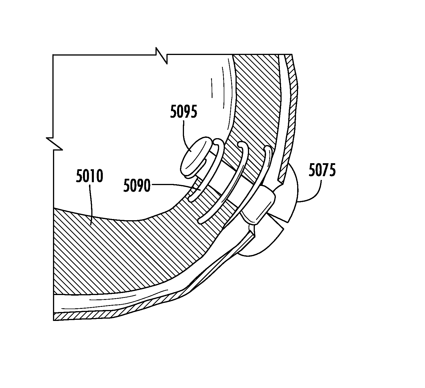
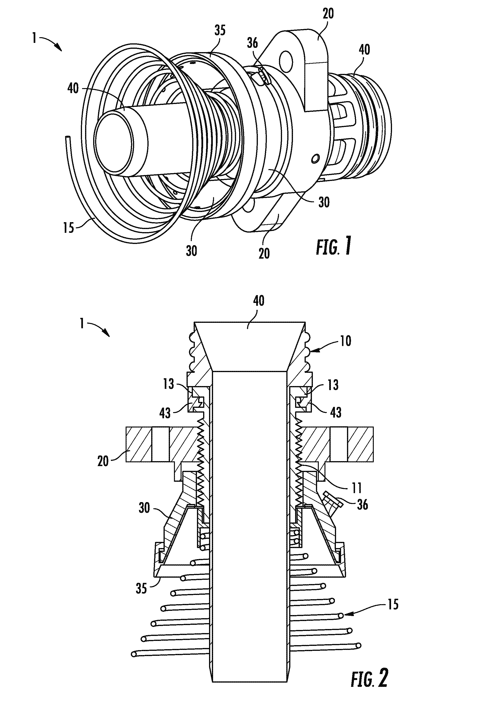
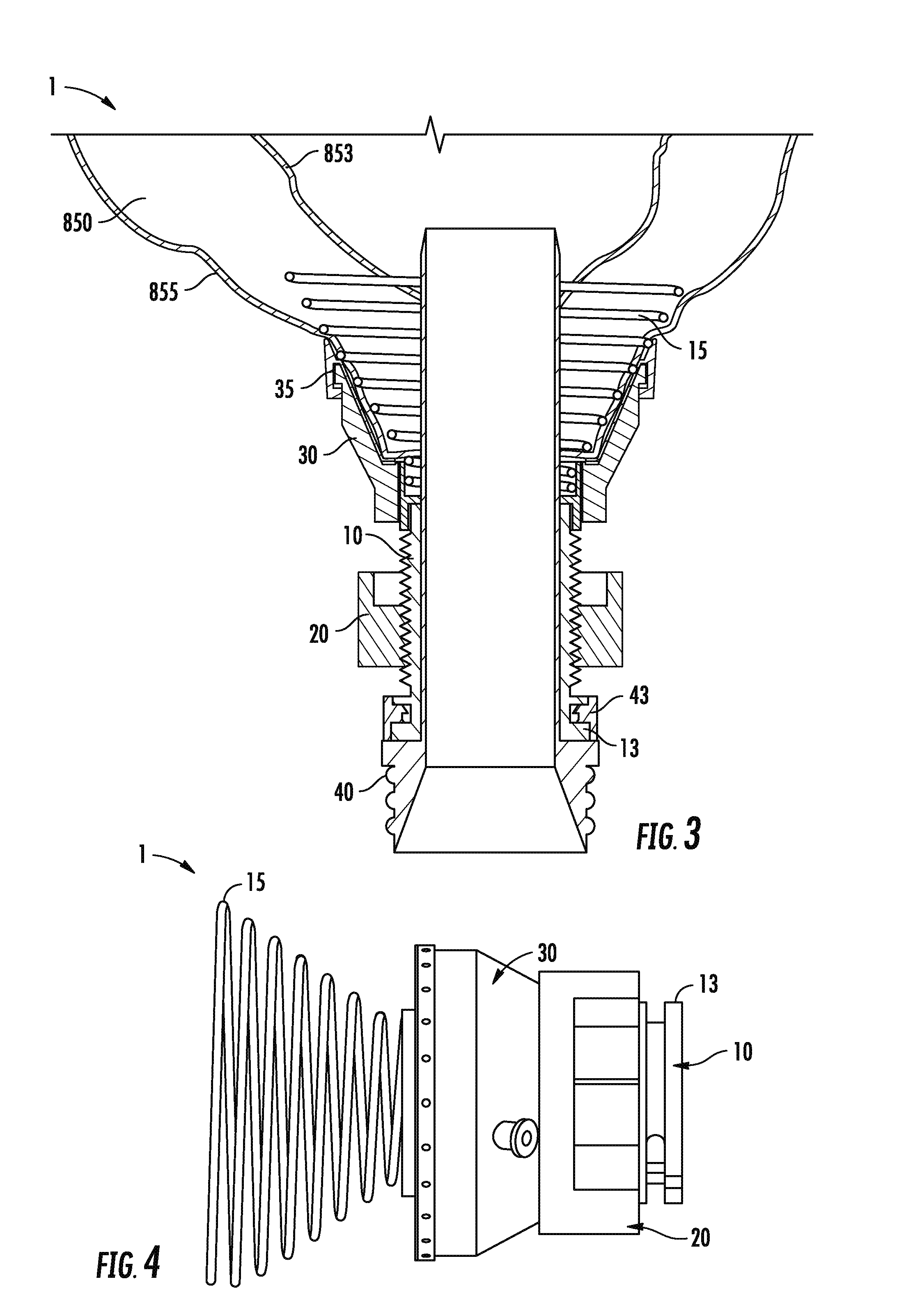
[0085]Certain embodiments of the invention provide devices, methods and systems for using a conduit device through a tissue wall of a patient, comprising: an outer conduit lumen; an inner conduit lumen adapted for insertion at least partially through the outer lumen; an attaching device in communication with a distal end of one of the outer lumen or the inner lumen, wherein the attaching device is ada...


PUM


Abstract

Systems and methods for accessing, stabilizing and sealing a device attached to a tissue surface comprising a tissue attaching device having an outer base ring defining an opening therethrough and a distally projecting tissue attachment element. The systems variously utilize annular sealing flanges distally attached to the outer base ring outside or inside the tissue attachment element to create a fluid tight seal. The systems variously utilize coils with regions of differing pitch to create sealing tissue pressure. Methods for installing an apical attaching device with a transapical port into a patient, comprising assessing the patient's viability for installation of the apical attaching device by determining an Index of Tissue Elasticity (ITE).

Description

CROSS REFERENCES TO RELATED APPLICATIONS[0001]This application is a continuation of PCT Application No. PCT / US2014 / 117087 filed Jan. 27, 2014 which claims priority to U.S. Provisional Application Ser. Nos. 61 / 756,885 filed Jan. 25, 2013, 61 / 777,796 filed Mar. 12, 2013, 61 / 819,237 filed May 3, 2013, and 61 / 835,712 filed Jun. 17, 2013, the entire contents of which are incorporated by reference herewith.FIELD OF THE INVENTION[0002]This invention relates to devices and methods for creating, maintaining, controlling, and closing a fluid communication between opposing surfaces of a tissue wall.BACKGROUND OF THE INVENTION[0003]In the human body, various organs contain fluids both in liquids and gaseous forms within tissue layers or cavities formed by tissue. These liquids may or may not be under pressure. The tissue walls around these cavities are normally designed to confine these liquids to specific areas of the body. Blood as in the heart and vasculature in order to preserve its volume ...

Claims


Application Information

Patent Timeline
Patent Type & Authority Applications(United States)
IPC IPC(8): A61B17/064A61B17/00A61B17/34
CPCA61B17/0644A61B2017/3425A61B17/3421A61B17/0057A61M39/06A61B2017/00247A61B2017/00252A61B2017/00305A61B2017/00318A61B2017/00349A61B2017/00575A61B2017/00588A61B2017/00615A61B2017/00623A61B2017/0649A61B17/0643A61B17/0401A61B2017/00336A61B2017/00703A61B2017/00867A61B2017/00902A61B2017/0406A61B2017/0464A61B2017/0498A61B2017/306A61B2017/3419A61B2017/3466A61B2017/3488A61B2090/0801A61B17/3423
Inventor JIMENEZ, JORGE HERNANWEST, DUSTIN SETHCUNNIFFE, BRENDANFEENEY, THOMASGREENE, JAMES L.STEPHENS, ROBIN
Owner APICA CARDIOVASCULAR
Why Eureka
Try Eureka