Radiography system and radiography method

a radiography system and radiography technology, applied in the field of radiography system and radiography method, can solve the problems of wasting time taken for image examination, difficult to perceive multiple diseased tissues or the like, and long time to complete the production of such a large number of tomographic images, so as to achieve the effect of completing the image examination in a shorter time and ensuring the accuracy of the resul

Active Publication Date: 2016-01-21
FUJIFILM CORP
View PDF3 Cites 12 Cited by
  • Summary
  • Abstract
  • Description
  • Claims
  • Application Information

AI Technical Summary

Benefits of technology

[0012]An object of the present invention is to provide a radiography system and a radiography method, which enable an exact image examination while reducing the time required therefor.
[0020]In the radiography system and the radiography method in accordance with the present invention, minimum tomographic images required for the image examination are selected to be produced and displayed preferentially according to the sweep angle from among the tomographic images to be produced for diagnosis, enabling completing the image examination in a shorter time while ensuring the precision thereof.

Problems solved by technology

Therefore, from a projection image of a subject (e.g. a patient for diagnosis), it is difficult to perceive multiple diseased tissues or the like, which may exist at different depths in the subject, distinguishably from each other.
However, for the tomosynthesis, it is necessary to produce several tens to more than one hundred tomographic images, and it takes a long time to complete production of such a great number of tomographic images.
The time waiting for the production of tomographic images is one of causes that waste time taken for the image examination.
This method may be effective to save the preparation time required for acquiring a set of projection images which are used for production of tomographic images, but could not reduce the time taken for the image examination.
However, as being produced using a reduced number of projection images, the incomplete tomographic image is inferior in quality to a tomographic image that is produced using all of the series of projection images.
Therefore, depending on the skills in imaging and examining the tomographic images, there may be cases where the radiologist cannot exactly determine whether the image acquisition is successful or not on the basis of the incomplete tomographic image.

Method used


Image

  • Radiography system and radiography method
  • Radiography system and radiography method
  • Radiography system and radiography method

Examples


Embodiment Construction

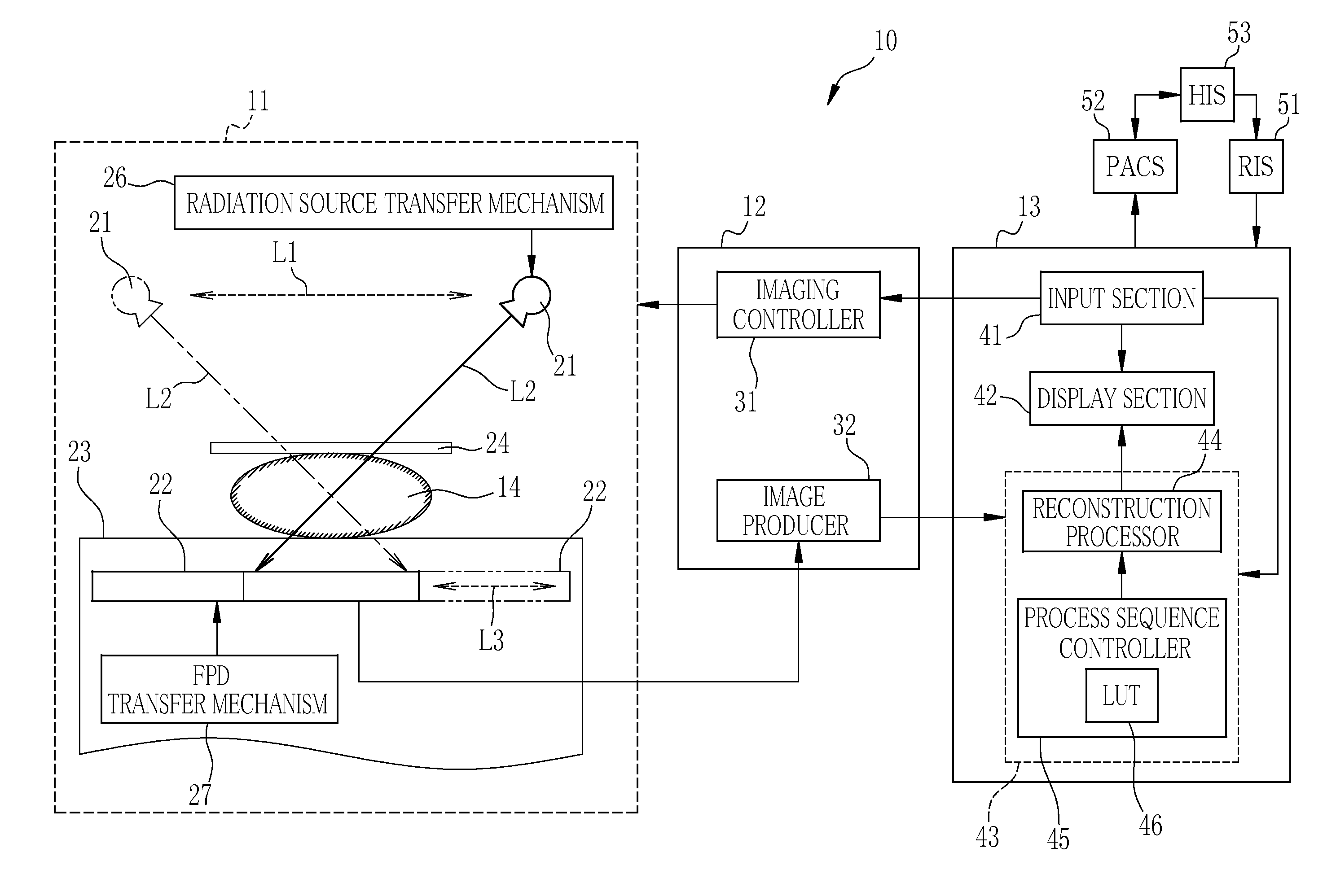
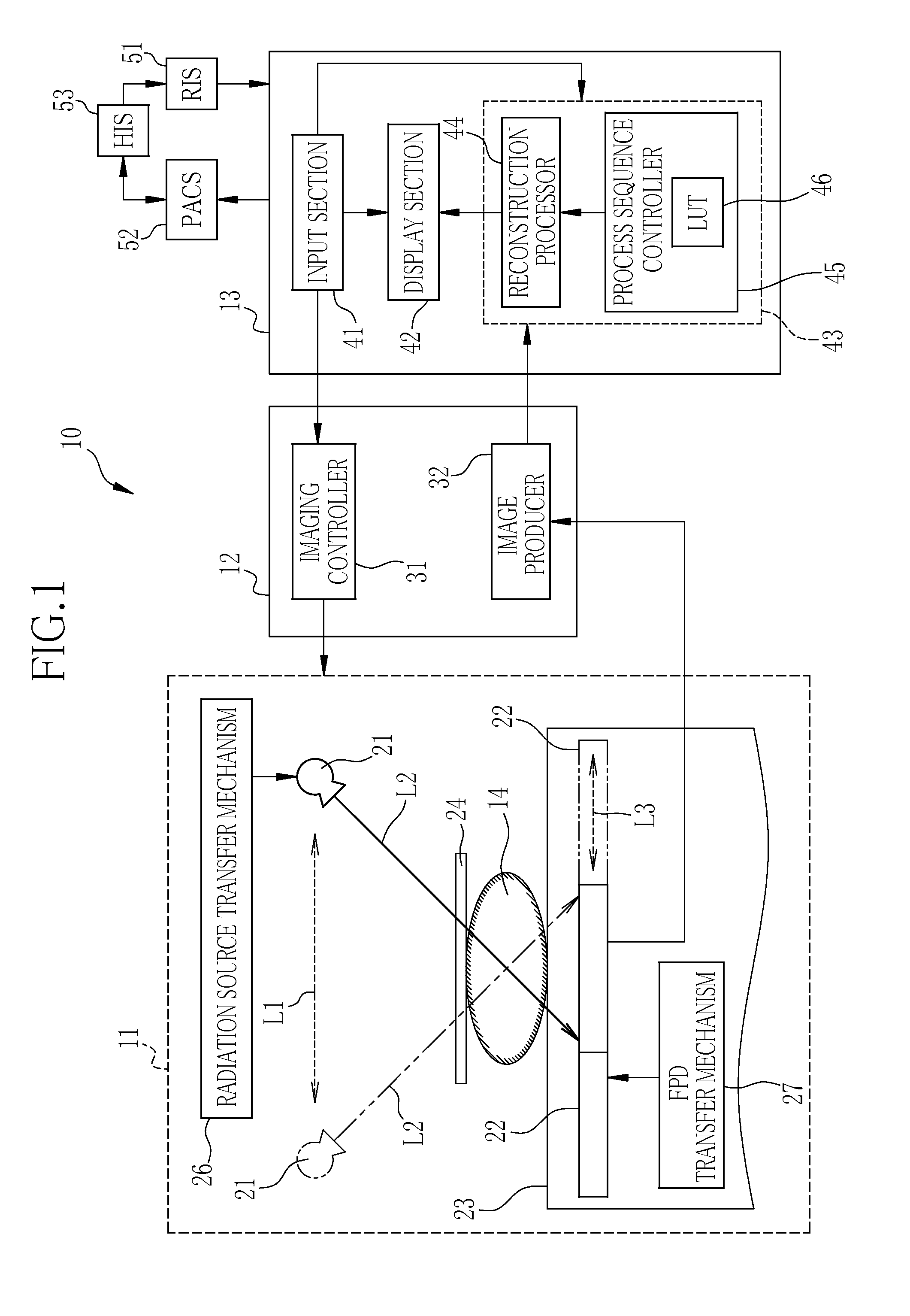
[0035]As shown in FIG. 1, a radiography system 10 is typically equipped with an imaging unit 11, a control unit 12 for controlling the imaging unit 11, and a console 13 for operating input / output of various data and the like between the control unit 12 and other external devices.

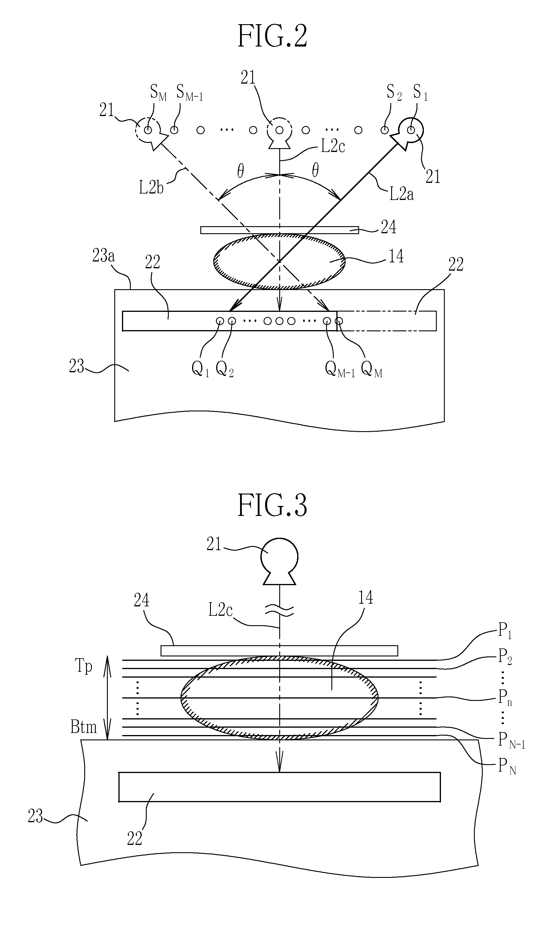
[0036]In this example, the imaging unit 11 is a digital mammography apparatus which is mainly provided with a radiation source 21, a flat panel detector (FPD) 22 as a radiographic image detector, an imaging stage 23 and a pressing plate 24, wherein a breast mass is an imaging subject 14. The subject 14 is set on the imaging stage 23 and pressed onto the imaging stage 23 by the pressing plate 24 which is movable relative to the imaging stage 23, so as to be imaged in a flatten-out condition.

[0037]The radiation source 21 generates X-rays. The quantity and quality of X-rays generated from the radiation source 21 are adjustable by means of tube voltage and tube current. The radiation source 21 is mounted to a ra...


PUM


Abstract

A radiography system includes an imaging controller, a reconstruction processor, a process sequence controller and a display section. The imaging controller controls a radiation source to move in a range of a designated sweep angle and project X-rays at different angles to an imaging subject, while controlling a flat panel detector to acquire a set of projection images. The reconstruction processor uses the set of projection images to produce a plurality of tomographic images for use in diagnosis. The process sequence controller determines a parameter for sorting the plurality of tomographic images into first and second groups according to the sweep angle of the radiation source so as the first group of tomographic images to be produced prior to the second group of tomographic images. The display section displays at least the first group of tomographic images.

Description

CROSS-REFERENCE TO RELATED APPLICATIONS[0001]This application is a Continuation of PCT International Application No. PCT / JP2014 / 057230 filed on Mar. 18, 2014, which claims priority under 35 U.S.C §119(a) to Japanese Patent Application No. 2013-073588 filed Mar. 29, 2013. The above application is hereby expressly incorporated by reference, in its entirety, into the present application.BACKGROUND OF THE INVENTION[0002]1. Field of the Invention[0003]The present invention relates to a radiography system and a radiography method, wherein tomographic images of a subject are obtained through reconstruction of radiographic images which are acquired from the subject by projecting radiation at different projection angles.[0004]2. Description Related to the Prior Art[0005]Radiography systems have been widely spread, which are each equipped with a radiation source for projecting radiation (e.g. x-rays) to a subject and a radiographic image detector for detecting radiographic images of the subje...

Claims


Application Information

Patent Timeline
Patent Type & Authority Applications(United States)
IPC IPC(8): A61B6/02A61B6/00
CPCA61B6/025A61B6/5205A61B6/54A61B6/463A61B6/0414A61B6/4233A61B6/502A61B6/547
Inventor MORITA, SEIKIHIGUCHI, TAKAAKISUGAHARA, MASATAKASASAKI, WATARU
Owner FUJIFILM CORP
Why Eureka
Try Eureka