Alpha connexin c-terminal (ACT) peptides for use in transplant

a technology of c-terminal and act peptides, which is applied in the field of alpha connexin c-terminal (act) peptides for transplantation, can solve problems such as cell injury

Active Publication Date: 2016-05-05
MUSC FOUND FOR RES DEV
View PDF0 Cites 9 Cited by
  • Summary
  • Abstract
  • Description
  • Claims
  • Application Information

AI Technical Summary

Benefits of technology

[0013]In one embodiment, the polypeptide promotes cell-cell communication. In another embodiment, the polypeptide stabilizes gap junctions in cells. In yet another embodiment, the polypeptide stabilizes tight junctions in cells. In one embodiment, the polypeptide mitigates hemichannel activity in cells. In one embodiment, the polypeptide inhibits apoptosis in cells. In another embodiment, the polypeptide inhibits mitochondrial oxidant production. In another embodiment, the polypeptide promotes the integrity of endothelial cells. In another embodiment, the polypeptide promotes barrier function of endothelial cells. In some embodiments, the cells are the cells of an organ for transplantation from a donor to a recipient.

Problems solved by technology

In one aspect, the cellular injury is caused by cold preservation induced damage.

Method used


Image

  • Alpha connexin c-terminal (ACT) peptides for use in transplant
  • Alpha connexin c-terminal (ACT) peptides for use in transplant
  • Alpha connexin c-terminal (ACT) peptides for use in transplant

Examples


example 1

1. Example 1

[0275]As shown in FIG. 1, the alpha connexin carboxy-terminal (ACT) polypeptide ACT1 prevents VEGF-induced deterioration of TER in ARPE-19 cells. Trans-epithelial resistance (TER) measurements, using ARPE19 cell (immortalized human RPE cells) monolayers revealed that VEGF leads to rapid deterioration, which was blocked by pre-treating the cells with the ACT peptide. Thus, while not wishing to be bound by theory, stabilizing the tight junction proteins with the ACT peptide can prevent loss of tight junction disintegration and thus damage to RPE / Bruch's membrane.

[0276]ACT1 Peptide contains an amino terminal cell internalization sequence. Together with a mild detergent that is used in ocular applications, Brij-78 the antenapedia sequence assists in permeation of ACT1 into interior fluids and tissues of the eye. In some aspects, the ability of ACT1 to enter the internal fluids and tissues of eye is a mode of action of ACT1 in treating diseases of the eye such as macular dege...

example 2

2. Example 2

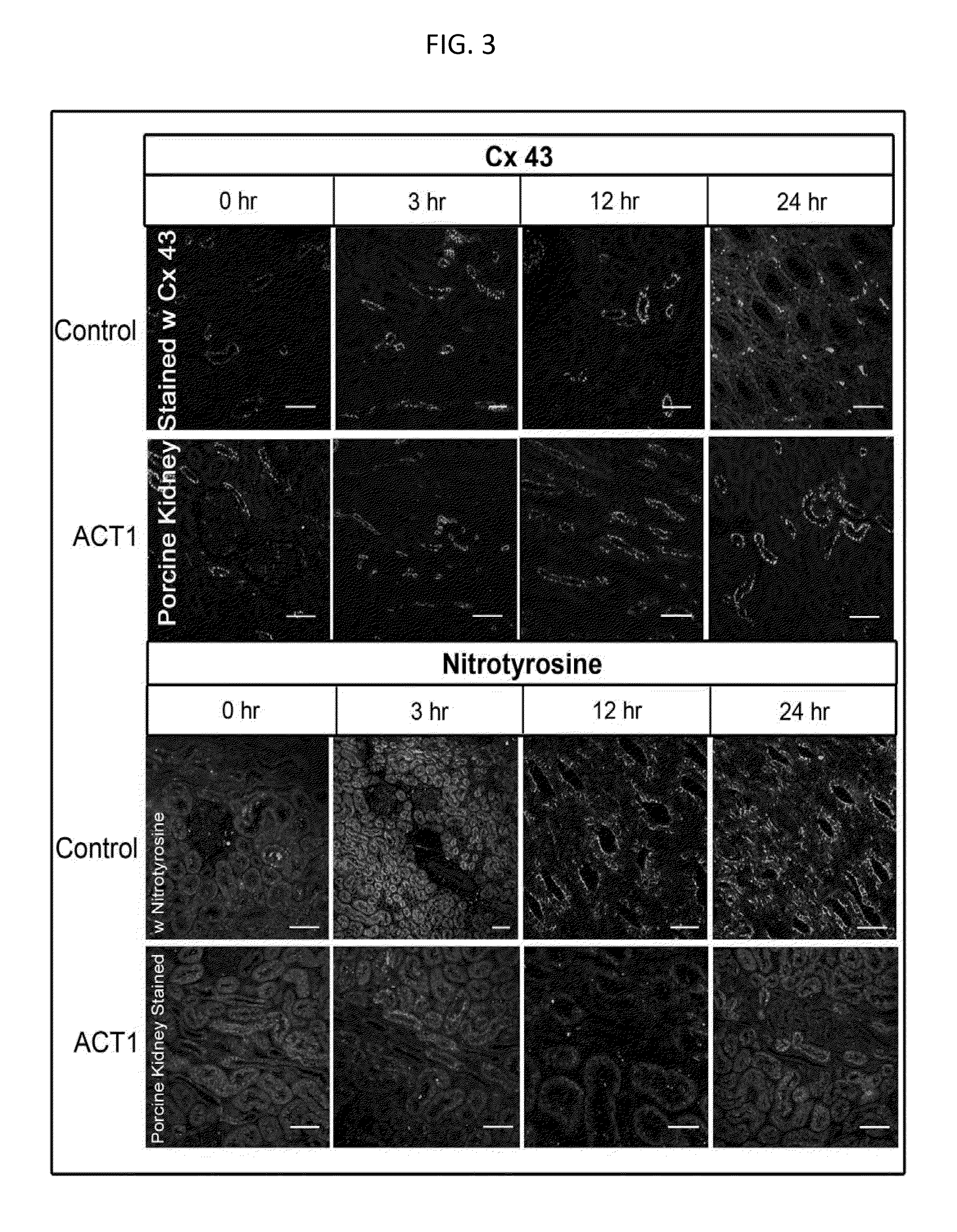
[0283]As shown in FIG. 3, ACT1 peptide stabilizes gap junctions (Cx43) and minimizes mitochondrial oxidant production (nitrotyrosine) and apoptosis (TUNEL and caspase) in porcine kidney models of cold ischemia.

[0284]i. Results

[0285]Punctate Cx43 staining in the membrane (gap junctions) were preserved in ACT1 peptide-treated kidneys and early control biopsies, while at 24 h Cx43 staining became more diffuse and appeared to localize to the cytoplasm of cells in the control kidneys. The 12 and 24 h sections demonstrated intense, localized nitrotyrosine staining in the apical and basolateral areas of control kidney cells in comparison to the ACT1 peptide-treated samples. There were no changes in nitrotyrosine staining in the presence of ACT1 peptide or in time zero control biopsies that were not subjected to cold ischemia. Apoptotic cells were also observed in the 24 h control (data not shown).

[0286]ii. Methods:

[0287]These studies were conducted using kidneys procured from 2...

example 3

3. Example 3

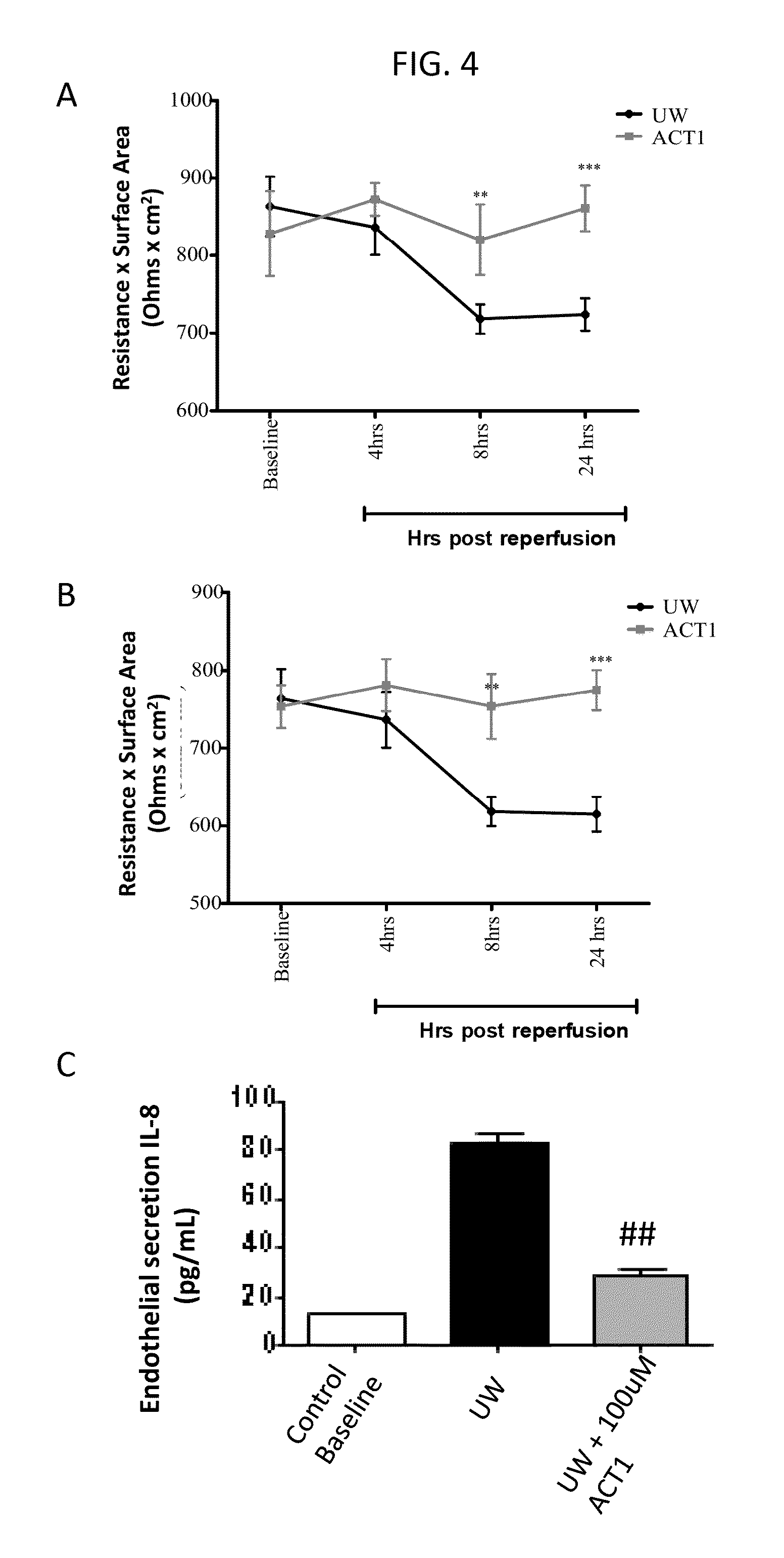
[0288]FIG. 4 shows the ability of ACT1 peptide to protect endothelial cells.

[0289]i. Results

[0290]Storage of both epithelial and endothelial cell with Belzer's University of Wisconsin (UW) solution containing 100 μM ACT1 peptide significantly reduced cellular injury as compared to untreated controls (FIGS. 4A and 4B). Further, supernatants and cell lysates were collected to measure IL-8 secretion and MHC II expression. Treatment of either cell type with UW solution supplemented with ACT1 peptide was associated with significant reduction in IL-8 secretion (FIG. 4C) and MHC Class II expression (endothelial cells, data not shown).

[0291]ii. Methods:

[0292]These studies were conducted using a modification of the in vitro donor cold storage and reperfusion injury model (Casiraghi et al., 2009). Human umbilical vein endothelial cells (HUVECs) and mouse microvascular endothelial cells were grown to confluence on transwells and transendothelial resistance (TEER) was recorded. To m...


PUM


Abstract

The present disclosure provides compositions and methods for treating or preventing macular degeneration in a subject. The disclosure also provides compositions and methods for preserving organs and tissues for transplantation, and for preventing cellular injury in organs or in subjects.

Description

CROSS-REFERENCE TO RELATED APPLICATIONS[0001]This application is a continuation-in-part of U.S. application Ser. No. 13 / 935,848, filed Jul. 5, 2013, which is a continuation of U.S. application Ser. No. 12 / 665,596, filed Jul. 21, 2010, which is the U.S. National Phase of International Application No. PCT / US08 / 67944, filed Jun. 23, 2008, which claims priority to U.S. Provisional Application Ser. No. 60 / 945,493, filed Jun. 21, 2007; and U.S. application Ser. No. 13 / 935,848 further claims priority as a continuation-in-part of U.S. application Ser. No. 13 / 842,506, filed Mar. 15, 2013, which is a continuation of U.S. application Ser. No. 13 / 715,626 filed Dec. 14, 2012, which is a continuation of U.S. application Ser. No. 12 / 871,461, filed Aug. 30, 2010, now U.S. Pat. No. 8,357,668, which is a divisional of U.S. application Ser. No. 11 / 721,529, filed Jun. 12, 2007, now U.S. Pat. No. 7,786,074, which is a U.S. National Phase of International Application No. PCT / US2005 / 046442, filed Dec. 20,...

Claims


Application Information

Patent Timeline
Patent Type & Authority Applications(United States)
IPC IPC(8): A01N1/02
CPCA01N1/0226
Inventor GHATNEKAR, GAUTAMGOURDIE, ROBERTJOURDAN, JANE
Owner MUSC FOUND FOR RES DEV
Why Eureka
Try Eureka