Systems and methods for analyzing medical images and creating a report

a technology of medical images and reports, applied in the field of systems and methods for analyzing medical images of tissue regions, can solve the problems of increasing the risk of breast and ovarian cancer, and achieve the effect of facilitating the comparison of patients and reducing lung function

Inactive Publication Date: 2016-07-14
IMBIO
View PDF2 Cites 71 Cited by
  • Summary
  • Abstract
  • Description
  • Claims
  • Application Information

AI Technical Summary

Benefits of technology

[0007]The present disclosure, in one or more embodiments, relates to a computer implemented method for assessing and communicating a patient's health status and risk. The method may include the steps of: receiving an imaging dataset of the patient, the imaging dataset comprising a plurality of voxels; automatically analyzing the imaging dataset for the presence and extent of an imaging biomarker; comparing the presence and extent of the imaging biomarker in the imaging dataset of the patient to the presence and extent of the imaging biomarker in historical imaging datasets previously acquired from other patients having known clinical outcomes; using the comparison to calculate personalized quantitative health status and risk metrics for the patient; and creating a report tailored for the intended user of the report to communicate the patient's personalized quantitative health status and risk metrics. In some embodiments the method may also include segmenting the imaging dataset of the patient into voxels corresponding to tissue of interest and voxels corresponding to tissue of no interest. In some embodiments, the imaging biomarker may be the number of voxels with intensity below a threshold. The threshold may be in the range −910 HU to −960 HU. In some embodiments, the quantitative health status and risk metrics may be related to one or any combination of: myocardial infarction, Chronic Obstructive Pulmonary Disease (COPD), emphysema, lung cancer, decreased lung function, COPD exacerbations, coronary artery disease, and stroke.
[0008]Additionally, the present disclosure, in one or more embodiments, relates to a computer implemented method for assessing and communicating a patient's health status and risk. The method may include the steps of: receiving at least one medical image of a patient, the at least one medical image comprising a plurality of voxels; directly comparing the at least one medical image to historical image data previously acquired from other patients having known clinical outcomes; using the comparison to calculate personalized quantitative health status and risk metrics for the patient; and creating a tailored report based on indications of characteristics of the user to communicate the patient's personalized quantitative health status and risk metrics. In some embodiments, the direct comparison may use an unsupervised machine-learning algorithm. In other embodiments, the direct comparison may use a model-based algorithm. The method may further include segmenting the at least one medical image into voxels corresponding to a tissue of interest and voxels not of interest. In some embodiments, the method may further include automatically analyzing the voxels of interest for the presence and extent of an imaging biomarker; and co...

Problems solved by technology

The presence of altered forms of these genes res...

Method used


Image

  • Systems and methods for analyzing medical images and creating a report
  • Systems and methods for analyzing medical images and creating a report
  • Systems and methods for analyzing medical images and creating a report

Examples


Embodiment Construction

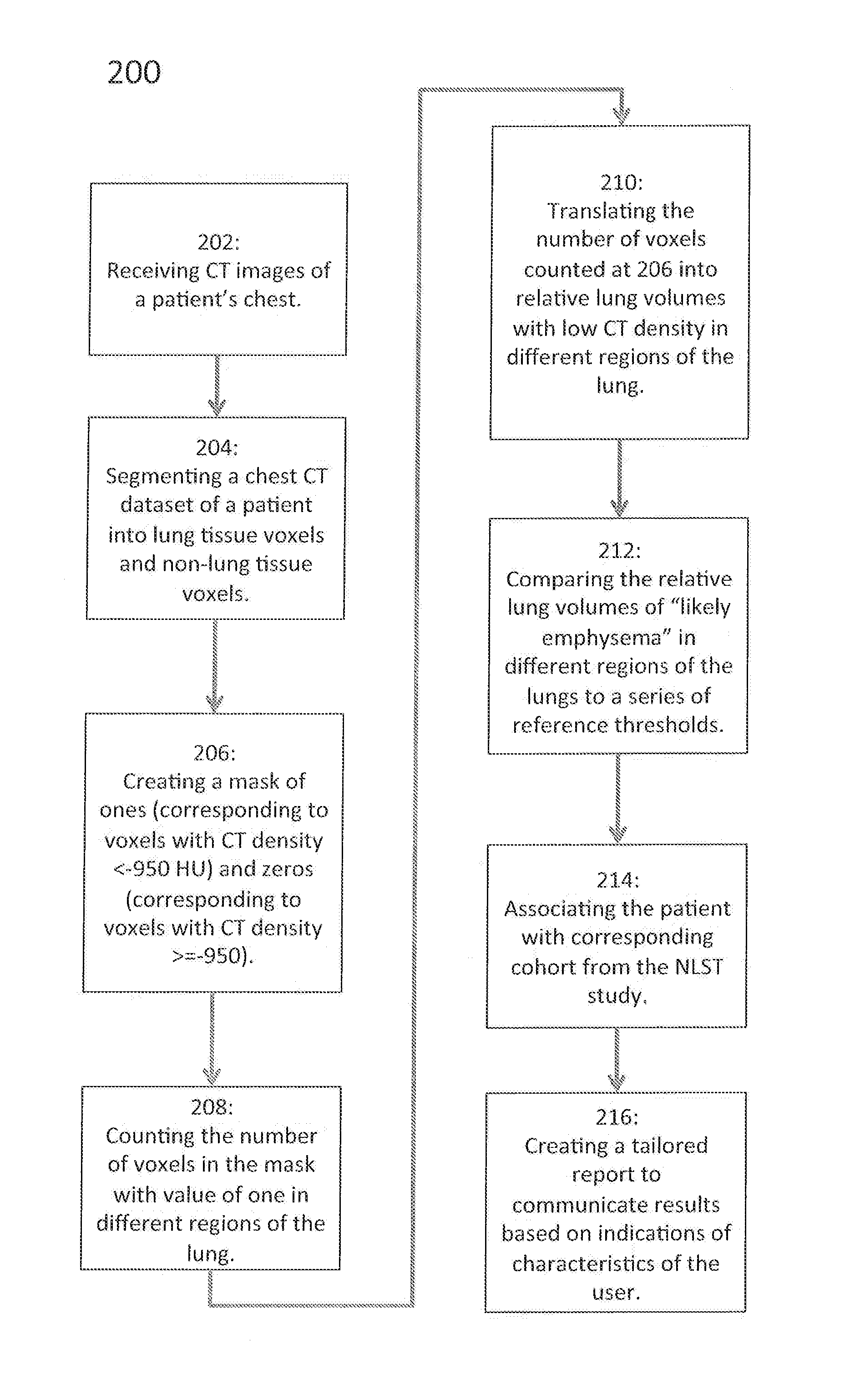
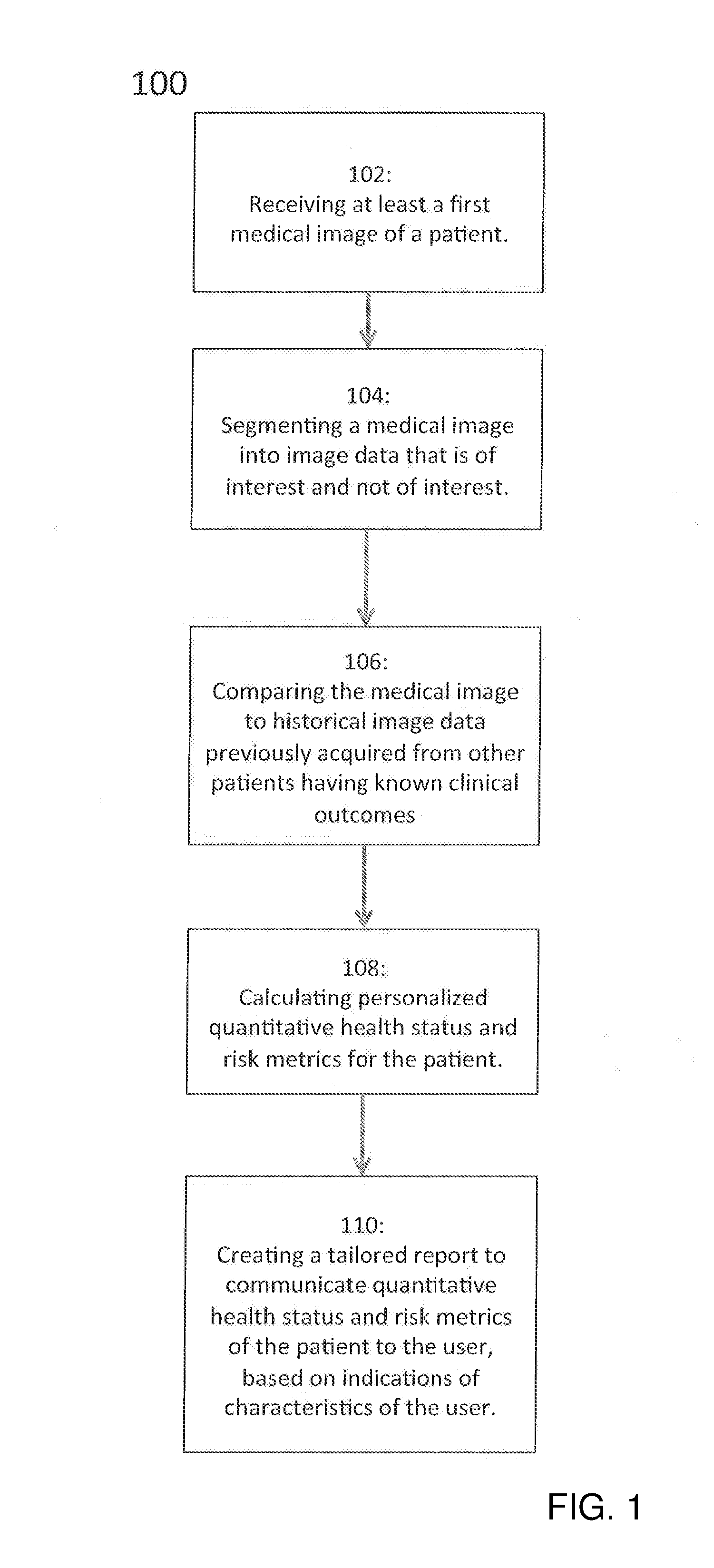
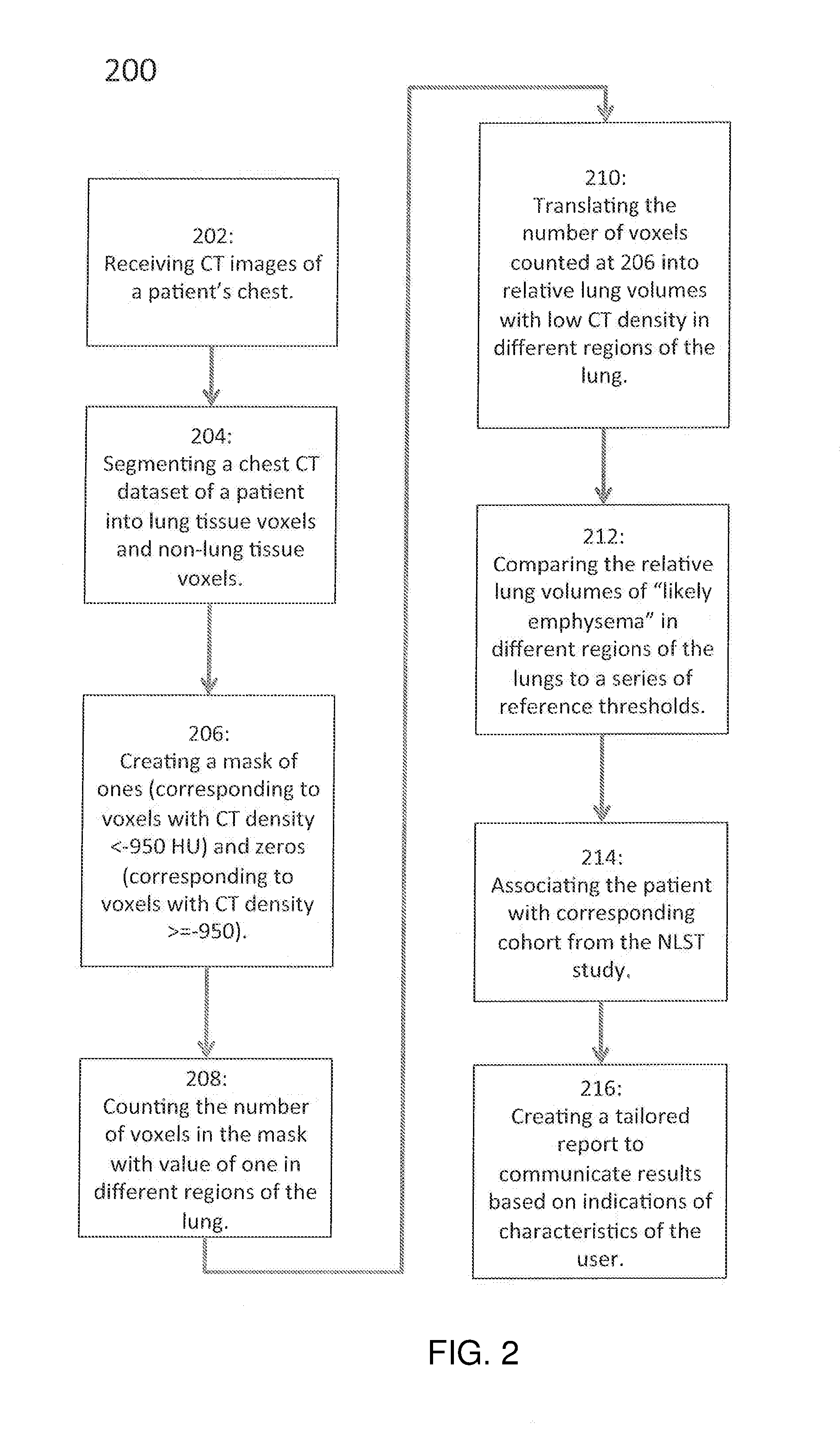
[0014]The present disclosure relates to computer-implemented systems and methods for automatically analyzing a patient's one or more medical images and creating at least one report that provides quantitative metrics related to the patient's current health status and their risks for future health outcomes. The analysis may be based on a computer-implemented algorithm that compares the patient's images to one or more comparison images of the same or similar tissue regions from one or more other individuals for whom the corresponding health status and / or outcomes are known. Multiple reports may be created for the same patient from the same medical images, wherein each of the multiple reports may be differently tailored for different types of intended users of the reports. As used herein, a “user” may be, for example, a patient, a patient's guardian or caregiver, a primary care physician, a nurse, a nurse practitioner, a chiropractor or osteopathic practitioner, a radiologist, a radiolo...


PUM


Abstract

Computer-implemented methods for automatically analyzing a patient's medical images and creating at least one report that provides quantitative metrics related to the patient's current health status and their risks for future health outcomes are provided. In at least one embodiment, the method comprises acquiring at least a first image from an imaging system; obtaining data based on the first image and a set of patient characteristic data; automatically analyzing the first image based on the patient characteristic data and displaying data in a user readable format.

Description

CROSS-REFERENCE TO RELATED APPLICATIONS[0001]This application claims priority to U.S. Provisional Application No. 62 / 101,167, entitled “Systems and Methods for Analyzing Medical Images and Creating a Report,” filed Jan. 8, 2015, the entirety of which is incorporated herein by reference.FIELD OF THE INVENTION[0002]The present disclosure relates to novel and advantageous systems and methods for analyzing medical images of tissue regions and, more particularly, to systems and methods for reporting information regarding the tissue regions to a patient, doctor, or other user.BACKGROUND OF THE INVENTION[0003]Medical imaging has been widely used to assess and monitor the status of various tissue regions within the body. For example, computed tomography (CT) and magnetic resonance imaging (MRI) are 3-dimensional, minimally invasive medical imaging techniques that are capable of providing high contrast images of tissue regions inside the body with excellent spatial resolution. Although these...

Claims


Application Information

Patent Timeline
IPC IPC(8): G06F19/00G16H15/00G16H30/40G16H50/30
CPCG06F19/321G06F19/345G06F19/3431G06T7/0016G06T2207/10072G06T2207/20081G06T2207/30048G06T2207/30061G16H50/30G16H50/20G16H15/00G16H30/40
Inventor MAIER, CYNTHIA F.AKGUN, CAN E.IVES, PHILIP S.KOJASOY, SHANNON
Owner IMBIO
Why Eureka
Try Eureka