Method and device for displaying medical images

a medical image and image technology, applied in the field of medical images, can solve the problems of difficult task of interpreting the information provided by tomosynthesis data, and achieve the effect of improving the assessment of disease development and improving viewing modalities

Active Publication Date: 2016-10-20
KONINKLJIJKE PHILIPS NV
View PDF1 Cites 16 Cited by
  • Summary
  • Abstract
  • Description
  • Claims
  • Application Information

AI Technical Summary

Benefits of technology

[0005]There may thus be a need to provide enhanced viewing modalities of a patient that enable an improved assessment of disease development.
[0012]The viewing of synthetic mammograms in oblique directions enables the presentation of the 3D tomosynthesis data in one or more views of a set of synthetic 2D mammograms as a first overview of the patient data, for example. This aids a radiologist in more effectively searching the 3D volume.
[0013]The use of a projection direction, which can be oblique, enables a radiologist to benefit from a range of potential views of the 3D data set, rather than using a view from a single, central projection only.
[0023]The viewing of synthetic mammograms in oblique directions enables the presentation of the 3D tomosynthesis data in one or more views of a series of synthetic 2D mammograms, for example as a first overview of the patient data. Hence, oblique synthetic mammograms support the radiologist in optimizing the workflow for reading a volumetric 3D tomosynthesis data set. Furthermore, a more accurate verification that an examination has been successful can be achieved with oblique synthetic mammograms at an acquisition workstation. Finally, it can be possible for a better alignment with prior mammograms to be achieved by choosing the most suitable angular direction to view the synthetic mammogram when that angle is oblique from a central acquisition direction.

Problems solved by technology

However, interpreting information provided from the tomosynthesis data is a challenging task, because not all information is clearly visible.

Method used


Image

  • Method and device for displaying medical images
  • Method and device for displaying medical images
  • Method and device for displaying medical images

Examples


Embodiment Construction

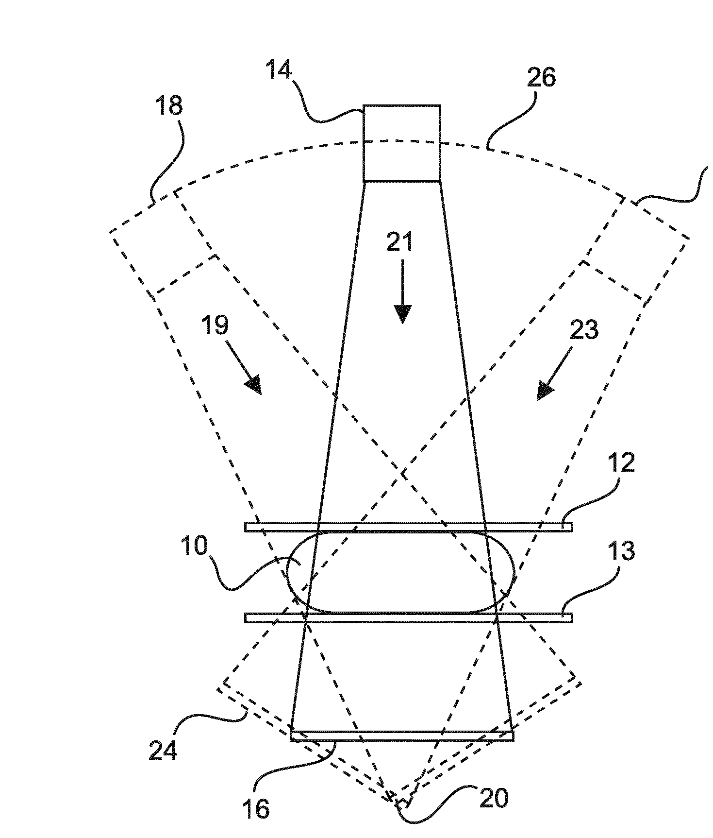
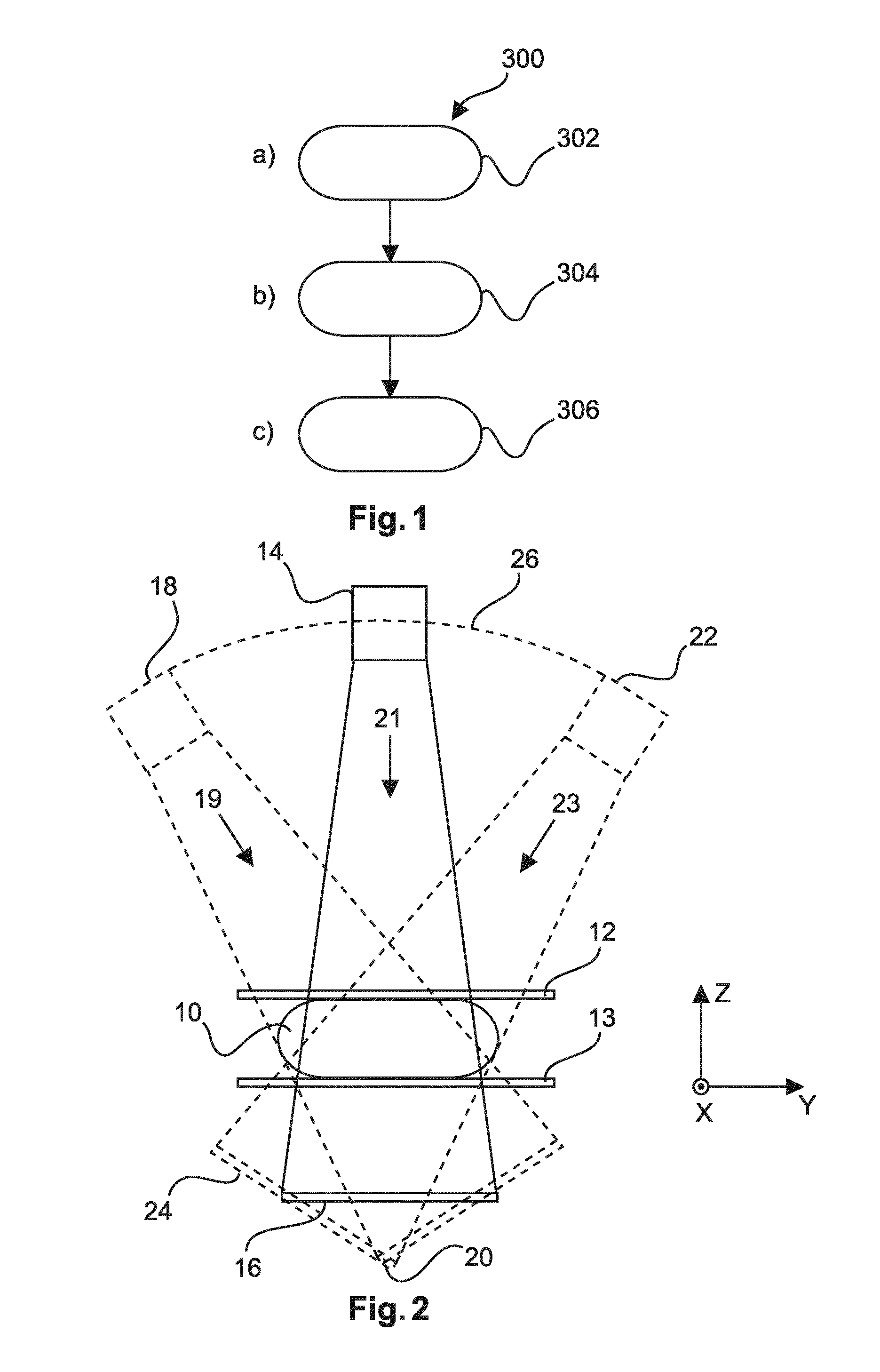
[0053]FIG. 1 shows a method 300 for displaying medical images acquired from a target using tomosynthesis. The method comprises the following steps: In a first step 302, processed 3D volume data representing a target, computed from a plurality of X-ray images through the target acquired along a plurality of acquisition directions is provided. In a second step 304, a first set of forward projections is computed in one or more projection directions through the target as represented by the processed 3D volume data, wherein at least one of the projection directions is oblique relative to a central acquisition plane. In a third step 306, the first set of forward projections is displayed as a corresponding set of synthetic mammograms.

[0054]The first step 302 is referred to as step a), the second step 304 is referred to as step b), and the third step 306 is referred to as step c).

[0055]According to the invention, the viewing of images from the 3D volume data, for example, synthetic mammogra...


PUM


Abstract

The present invention relates medical imaging, more particularly to the display of medical images acquired in a tomosynthesis process. Processed 3D volume data representing a target acquired from scanning may be viewed from a set of forward projection directions, in which one or more forward projections through the processed 3D volume data is oblique relative to a central acquisition direction of the scanning process. The first set of forward projections is then displayed as a set of synthetic mammograms. Therefore, a medical practitioner can more easily identify abnormalities within a target volume, because the relative displacement of abnormalities at different depths in the target volume will change at different rates dependent on the viewing angle as the viewing position is moved around the target.

Description

FIELD OF THE INVENTION[0001]The present invention relates to a method for displaying medical images. In particular, the invention relates to a device for displaying medical images acquired from a target using X-rays. The present invention also relates to a method for displaying medical images acquired from a target using X-rays, to an X-ray imaging system and to a computer program element and a computer-readable medium.BACKGROUND OF THE INVENTION[0002]Mammogram screening, for example, is an important procedure for the detection and monitoring of breast abnormalities and diseases at an early stage. Medical personnel, such as doctors or radiologists, may detect abnormalities directly from X-ray images. The X-ray tomosynthesis method, for example, is growing in use. X-ray tomosynthesis is a technique for producing 3D image volumes through a target using X-ray systems, although other techniques for providing such 3D volumes are known.[0003]U.S. Pat. No. 8,044,972 discusses a system and ...

Claims


Application Information

Patent Timeline
Patent Type & Authority Applications(United States)
IPC IPC(8): A61B6/00A61B6/02
CPCA61B6/466A61B6/025A61B6/5223A61B6/4452A61B6/467A61B6/502G06T11/008
Inventor ERHARD, KLAUSHOMANN, HANNO HEYKEREHN, JONAS RIKARD
Owner KONINKLJIJKE PHILIPS NV
Why Eureka
Try Eureka