
Methods for inducing electroporation and tissue ablation

a tissue ablation and electroporation technology, applied in the field of electroporation and tissue ablation, can solve the problems of increasing complexity and realism, ineffective and incomplete purely experimental approaches, and poorly understood electroporation field stimulation of cells, and achieves low energy permeability, effective induces cytosolic components, and high permeability state

Inactive Publication Date: 2016-12-15
VIRGINIA TECH INTPROP INC +1
View PDF6 Cites 61 Cited by
  • Summary
  • Abstract
  • Description
  • Claims
  • Application Information

AI Technical Summary

Benefits of technology

The present invention is about a new way to make holes in cells using electricity. The invention uses a method called electroporation, which involves using a single electric charge to disrupt the cell membrane and release important parts of the cell. This method uses less energy and less heat than other methods, which can damage the cells being treated. Overall, this invention provides a way to make holes in cells with reduced risk of damage.

Problems solved by technology

In spite of this progress, however, electromagnetic field stimulation of cells remains poorly understood.
Purely experimental approaches are inefficient and incomplete, because the combined cellular / field parameter space is huge.
This motivates the pursuit of multiscale models with increasing complexity and realism.
This may result in inadvertent thermal damage because when such intense electric fields are applied, excessive electrical current may pass through the resistive tissue causing unwanted heating.
However, there are challenges associated with the use of IRE for tumor ablation.
For example, ablation of large volumes of tissue with IRE remains difficult because the larger electric fields (for example, greater than 2500 V / cm) that would create larger lesions may also damage surrounding nerves and the cardiovascular system (Jiang et al., 2015).
In addition, some studies have shown that incomplete treatment can result after IRE, possibly resulting in tumor recurrence (Jiang et al., 2015).

Method used


Image

  • Methods for inducing electroporation and tissue ablation
  • Methods for inducing electroporation and tissue ablation
  • Methods for inducing electroporation and tissue ablation

Examples


example 1

Long Lived Pores (LLP) and the High Permeability State

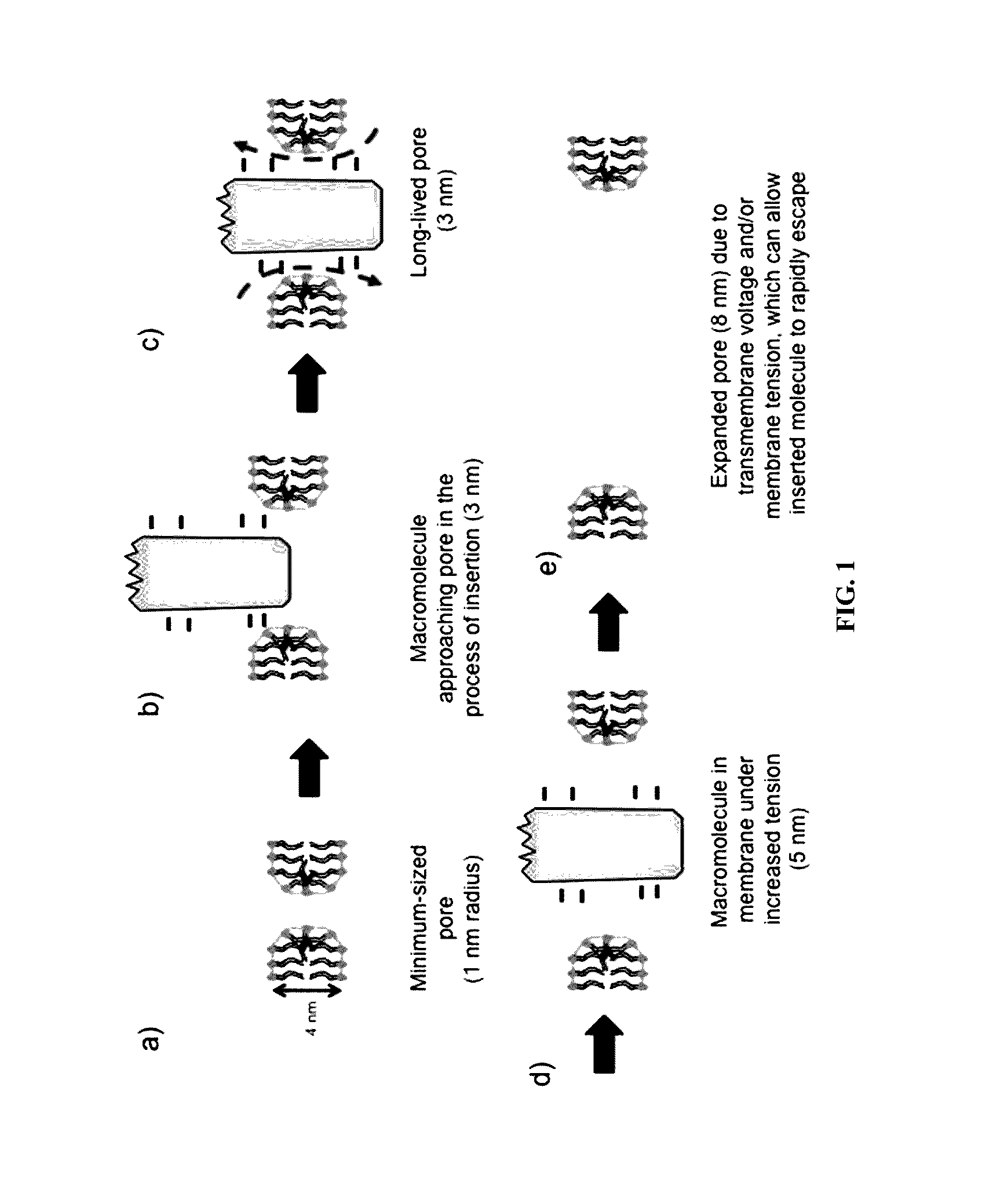
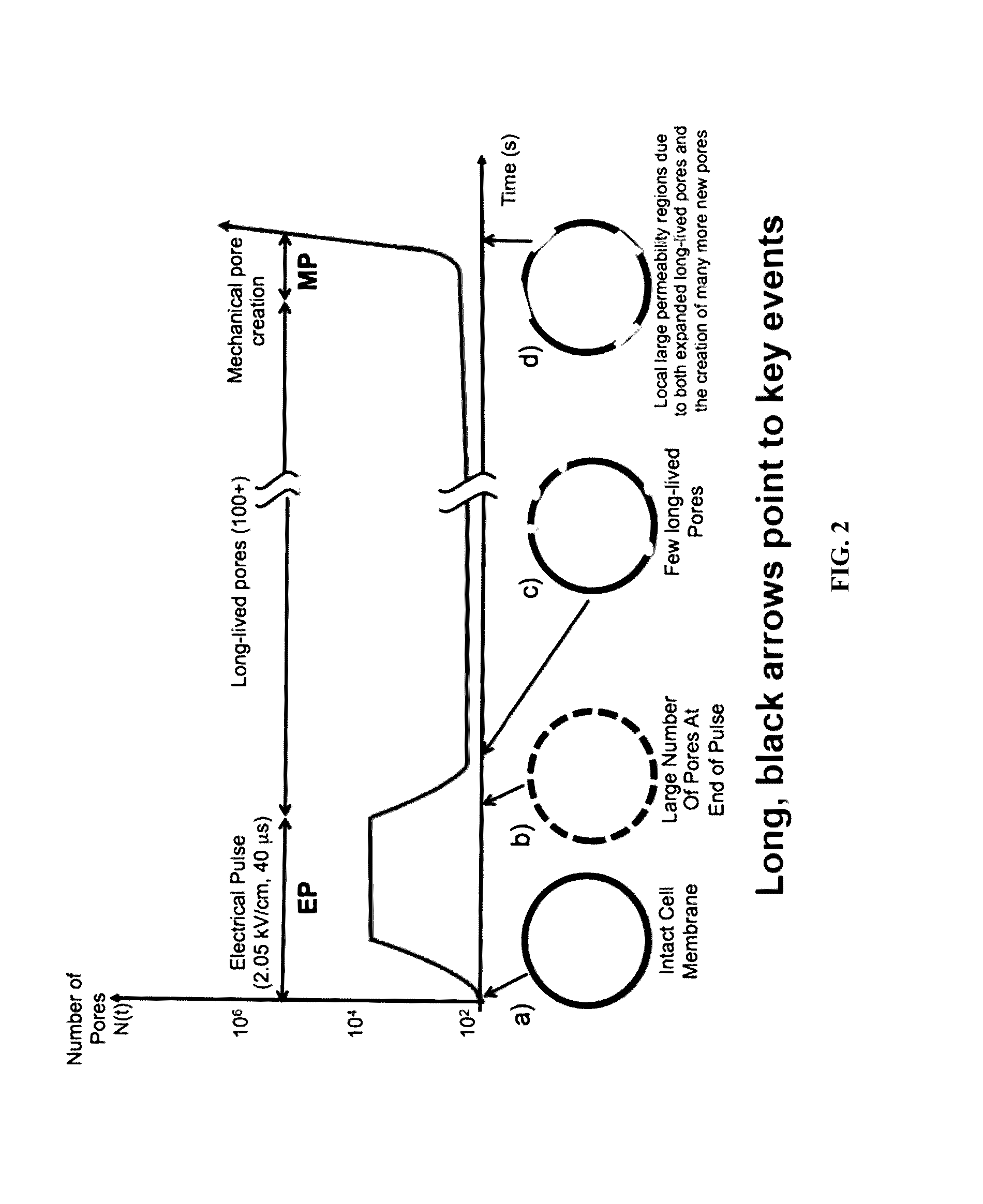
[0083]The high permeability state involves three phases of poration and involves exploiting intra- / extracellular osmotic pressure differences so that the electrical stimulus (and heating effects) can be smaller, and the change in permeability can be large. A model for the high permeability state is shown in FIG. 2. LLPs are involved as they allow EP to trigger mechanoporation (MP). The conceptual model is supported by quantitative simulations using an approximate cell model that includes dynamic EP with both TPs (traditional transient pores) and the LLPs. The initial simulations support the complex sequence of:

[0084]Phase 1: 40 microsecond EP pulse

[0085]Phase 2: Intervening time in which most TPs vanish, and about 100 LLPs survive. These LLPs supply / remove Na+, K+ and Cl− ions, causing a change in the cell osmotic pressure difference.

[0086]Phase 3: After some time, there is a nonlinear acceleration in LLP expansion, and then new ...

example 2

Cells can be Electroporated Using a Single Electrical Pulse

Methods

1. Cell Treatments in Microfluidic Chambers

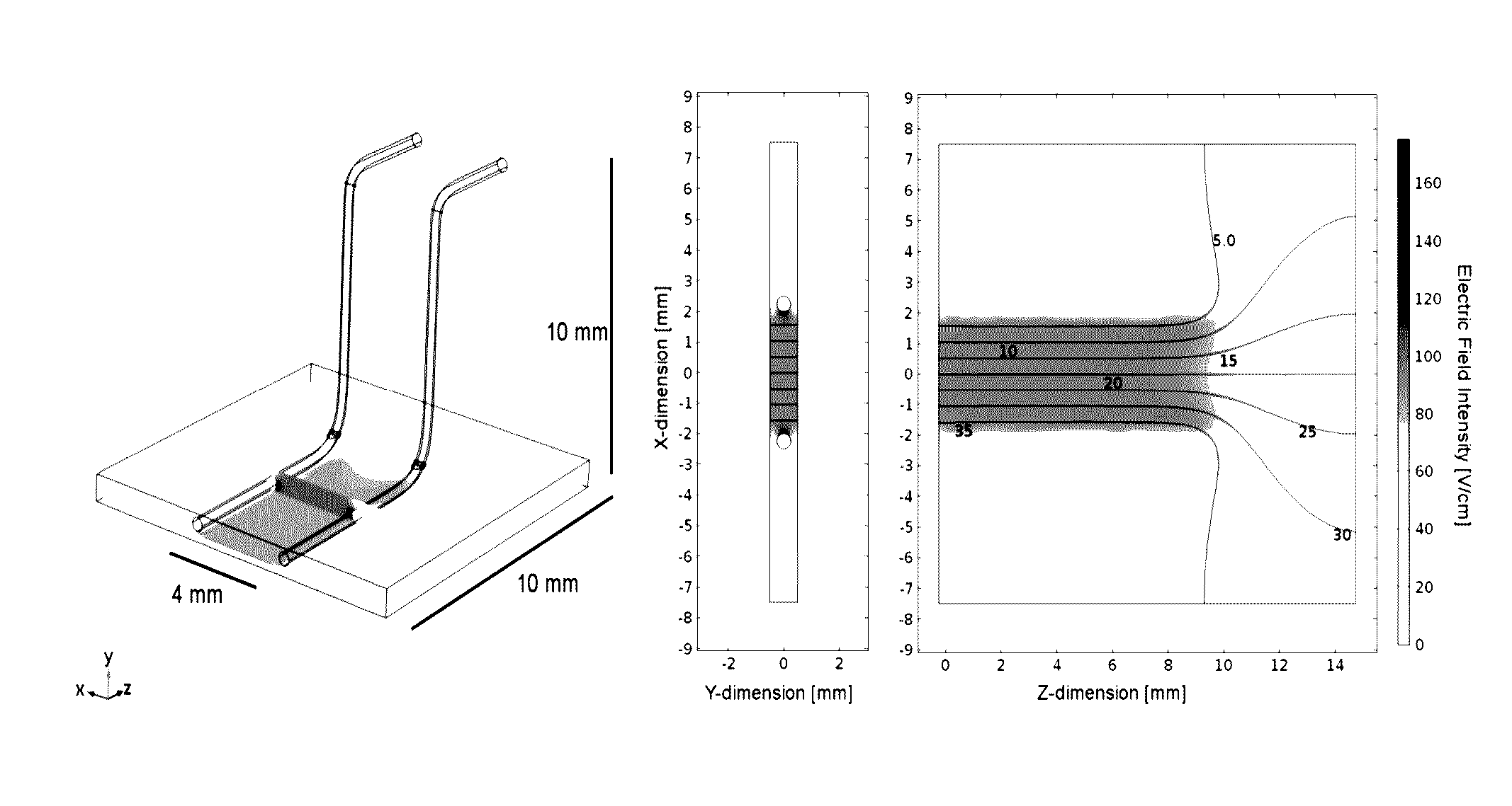
[0128]Data were obtained from two experimental setups: in a microfluidic device and in a growth chamber. Within the microfluidic device, Chinese hamster ovarian (CHO) cells were seeded at a density between 2-5×10⁶ cells / mL inside a microfluidic chip and allowed to adhere overnight. The channel height is approximately 90 μm and tapered along its length (approximately 3-4 cm) to generate a continuous electric field gradient across the length of the channel (FIG. 11). It was observed that a cell leakage event (FIG. 12) occurred over time and that it always preceded a large fluorescent intensification when it occurred. When quantified for each treatment, these leakage events occurred with increasing frequency as the electric field intensity was increased, above a certain threshold (FIG. 12). The observed leakage events occur differently, even under similar treatment times, using ...


PUM


Abstract

The invention encompasses a method of inducing a high permeability state in a cell membrane and a method for ablating a target tissue wherein the method comprises applying an electroporation pulse to a cell, wherein at a time after the electroporation pulse is applied, a plurality of long lived pores (LLPs) are formed in the cell membrane and the presence of the LLPs causes a change in the cell osmotic pressure difference. The invention also encompasses a method for ablating a target tissue using an electrical pulse regime that induces cell permeabilization and cell death, wherein the primary mechanism of cell death is as a result of electroporation.

Description

RELATED APPLICATION[0001]This application claims the benefit of U.S. Provisional Application 62 / 174,118, filed Jun. 11, 2015. The entire teaching of the above application is incorporated herein by reference.GOVERNMENT SUPPORT[0002]This invention was made with Government support under Grant No. R01 GM063857 awarded by the National Institutes of Health. The Government has certain rights in the invention.BACKGROUND OF THE INVENTION[0003]Cell membrane nanopores have been demonstrated experimentally using nanosecond electric field pulses14-17 in addition to conventional electroporation (EP). In spite of this progress, however, electromagnetic field stimulation of cells remains poorly understood. Purely experimental approaches are inefficient and incomplete, because the combined cellular / field parameter space is huge. This motivates the pursuit of multiscale models with increasing complexity and realism. Models offer objective guidance and perspective for investigators, and can provide ra...

Claims


Application Information

Patent Timeline
Patent Type & Authority Applications(United States)
IPC IPC(8): A61B18/14A61N1/30
CPCA61B18/1402A61N1/303A61B2018/00577A61B2018/1467A61B2018/00547A61B2018/00982A61B2018/00613A61N1/0412A61B18/1477A61N1/327
Inventor WEAVER, JAMES C.SON, REUBEN S.GOWRISHANKAR, THIRUVALLUR R.SWEENEY, DANIEL C.DAVALOS, RAFAEL V.
Owner VIRGINIA TECH INTPROP INC
Learn More